Pulmonary Emphysema Modeling & Pharmacodynamics Services
Introduction
Pulmonary emphysema, caused by damage to the air sacs in the lungs, is characterized by the gradual enlargement and destruction of alveoli in the lung, accompanied by destruction of their walls without obvious fibrosis. It is one of the lung conditions that causes breathing difficulties. Lung emphysema can progress to chronic obstructive pulmonary disease (COPD). This condition has a complex pathogenesis closely linked to factors such as smoking, infections, genetic mutations, and air pollution. Its typical pathological state involves decreased airway elasticity, over-distention, and hyperinflation of the terminal bronchioles, accompanied by airway wall destruction. With the increasing incidence and mortality rates of COPD, the treatment of emphysema has become a prominent area of research.
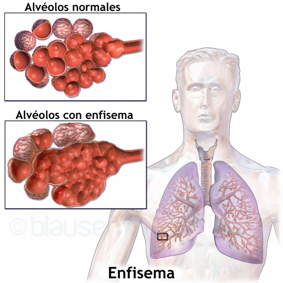 Fig.1 Normal versus emphysematous alveoli.
Available Pulmonary Emphysema Models
Creative Biolabs has established verified models to simulate the pathological characteristics of emphysema. We also have experience in evaluating pharmacology and pharmacodynamics of various drugs, such as small molecules, biopharmaceuticals, gene therapies, and cell therapies.
| Pulmonary Emphysema Models | Clinical Relevance | Primary Research Applications | Animal Species |
| PPE-Induced Pulmonary Emphysema Models | Accurately mimics emphysema associated with α1-antitrypsin deficiency (α1-ATD) in humans. | Novel Elastase Inhibitors, Antiprotease Therapies (e.g., α1-Antitrypsin Augmentation Therapy), and Cytoprotective Agents. | Mouse, Rat |
| Human Neutrophil Elastase (HNE) Model | Simulates lung tissue damage resulting from excessive release of neutrophil elastase. | Human Neutrophil Elastase (HNE) Specific Inhibitors (e.g., Sivelestat), and novel small or large molecule inhibitors designed for high clinical translatability. | Mouse, Hamster |
Evaluation Platform
We utilize advanced detection platforms to ensure stable and accurate results. For this purpose, we offer the following specialized platforms:
- Histopathology: Visualize alveolar destruction, inflammation, and fibrosis; quantify emphysema severity.
- Immunology: Validate mechanistic pathways, localize immune cell infiltration, and assess epithelial cell damage in lung tissue.
- Small animal in vivo imaging: Quantify lung volume, airspace enlargement, and emphysema progression; Offers non-invasive, 3D visualization of lung structure; longitudinal monitoring of disease progression.
- Cell and Molecular Biology: Utilizes in vitro models such as alveolar epithelial cells exposed to CSE/elastase, 3D cell cultures, and gene editing cells, to evaluate the activity or efficacy of the drug.
Applications
- Disease Modeling: They collectively validate the protease-antiprotease imbalance as a central mechanism in emphysema, providing a precise simulation of lung structural destruction.
- Mechanistic Studies: The HNE model induces lesion formation more rapidly, making it suitable for acute mechanistic studies. In contrast, the PPE model is better suited for simulating chronic, progressive lesions.
- Pharmacodynamic (PD) assessment: The models are widely employed for PD assessment of novel elastase inhibitors and anti-inflammatory drugs, accelerating the preclinical development of therapeutics with high translational value for Pulmonary Emphysema.
Our advantages
- Advanced Animal Models: We have developed two distinct animal models based on the clinical characteristics of emphysema. Additionally, our company is actively developing new models and can provide customized services to meet your specific research requirements.
- Integrated Detection Platforms: Our approach to confirming successful animal model development involves assessing inflammatory factors, monitoring animal status, and conducting lung tissue pathology. We also integrate advanced detection methods like proteomics and genomics to precisely measure pharmacodynamic indicators and perform in-depth mechanistic analysis.
- Professional Technical Team: Our experimental staff adheres to SOPs for all animal studies. Our team also includes scientists and statisticians with over 10 years of industry experience.
- One-Stop Service: Beyond pulmonary emphysema models, we offer a comprehensive range of other model services, including those for oncology, metabolic diseases, and autoimmune disorders.
Work with Us
- Summarize the project requirements and fill in the information collection form.
- Sign a CDA from both parties to further communicate information, such as targets.
- Select an animal model, discuss experimental design, and determine assay parameters.
- Project costing and project schedule forecasting.
- We provide a detailed project plan, including the required sample quantities, methods, and protocols.
- Both parties confirm the project details and start the project.
- Confirm the timeline of the project.
- We provide periodic results and information on the animal's condition.
- We will work together to make project adjustments as necessary.
- We provide a comprehensive project report promptly.
- We arrange transportation for the produced samples.
- We provide a discussion of the project results and help to arrange the next steps.
- Data storage and archiving.
FAQs
-
Q: How do you select the optimal model for a specific research question?
A: We align model choice with study goals, such as elastase models for protease-driven pathology and smoke models for chronic inflammation.
-
Q: Can you customize models to include comorbidities?
A: Yes, we develop combinatorial models via hypoxia exposure or vascular endothelial injury protocols.
-
Q: What safeguards can ensure model success rates?
A: Pilot studies validate induction parameters, such as elastase concentration and smoke exposure duration, prior to large-scale experiments.
-
Q: How is data scientific rigor maintained?
A: Rigorous data statistical analysis, randomized animal allocation, and stringent positive/negative controls are mandatory.
-
Q: What animal species do you use for emphysema models?
A: Mice (C57BL/6, BALB/c) are standard for cost-effectiveness and genetic manipulability, while rats or guinea pigs are available for larger airway or lung volume studies.
-
Q: What is the typical study duration for a chronic CSE model?
A: Study duration 8-12 weeks for mice (5 days/week exposure) to induce significant alveolar destruction; shorter (6-week) acute models for inflammation-focused studies.
-
Q: Do you provide raw data and statistical analysis?
A: Yes. Deliverables include raw data such as PFT traces, Western blots, quantified results, such as MLI, cytokine levels, and a detailed report with statistical tests (p-values, 95% CI).
-
Q: Can you model α1-antitrypsin deficiency (AATD) emphysema?
A: Yes, using SERPINA1⁻/⁻ knockout mice or human AAT-deficient serum transfer models to recapitulate genetic emphysema.
Published Data
This case highlights the use of the PPE-induced model and its application potential in pulmonary emphysema drug R&D. In the model group, significant alveolar septal thickening, alveolar dilation, and inflammatory cell infiltration were observed. Following treatment with Oxymatrine Liposomes (OMT-L), the destruction of alveolar structures was reduced, with alveolar morphology closely resembling that of the normal control group.
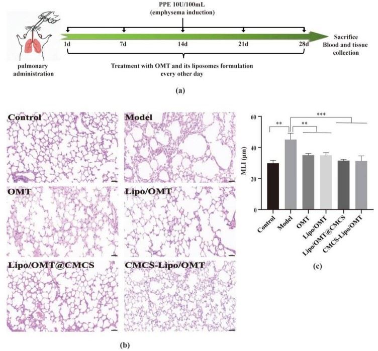 Fig.2 Representative histological images of lung sections from each group, stained with H&E. 1
Reference
- Peng, Jianqing et al. "Carboxymethyl Chitosan Modified Oxymatrine Liposomes for the Alleviation of Emphysema in Mice via Pulmonary Administration." Molecules (Basel, Switzerland) vol. 27,11 3610. https://doi.org/10.3390/molecules27113610. Distributed under Open Access license CC BY 4.0, without modification.
For Research Use Only.