Hi-Affi™ hPD-1/hPD-L1/hPD-L2 Triple Humanized Mouse Model
The combination of PD-1 protein and its ligands, including PD-L1 or PD-L2 will make T cells incapable and cannot effectively kill tumor cells. Thus, blocking the PD-1/PD-L1 and PD-1/PD-L2 pathways could relieve the suppression of T cells and improve the anti-tumor activity, which represents a strategy for cancer treatment. Creative Biolabs has successfully established an optimized Hi-Affi™ “humanized” animal platform to offer specialty manipulated hPD-1/hPD-L1/hPD-L2 triple humanized mice for our clients all over the world.
hPD-1/hPD-L1/hPD-L2 Molecule
Human programmed cell death protein-1 (hPD-1) is a member of the CD28 family and is an important immunosuppressive molecule on activated T cells, B cells, and monocytes. Its ligands are human programmed cell death ligand -1 (hPD-L1) and hPD-L2, which are highly homologous to B7-1 and B7-2 structures. Compared to hPD-L1, the expression range of hPD-L2 is relatively narrow. hPD-L1 is expressed on immune cells and tumor cells, while PD-L2 is mainly expressed on immune cells. The binding of both molecules to hPD-1 can inhibit T cell proliferation and cytokine production, and participate in the formation of peripheral autoimmunity tolerance.
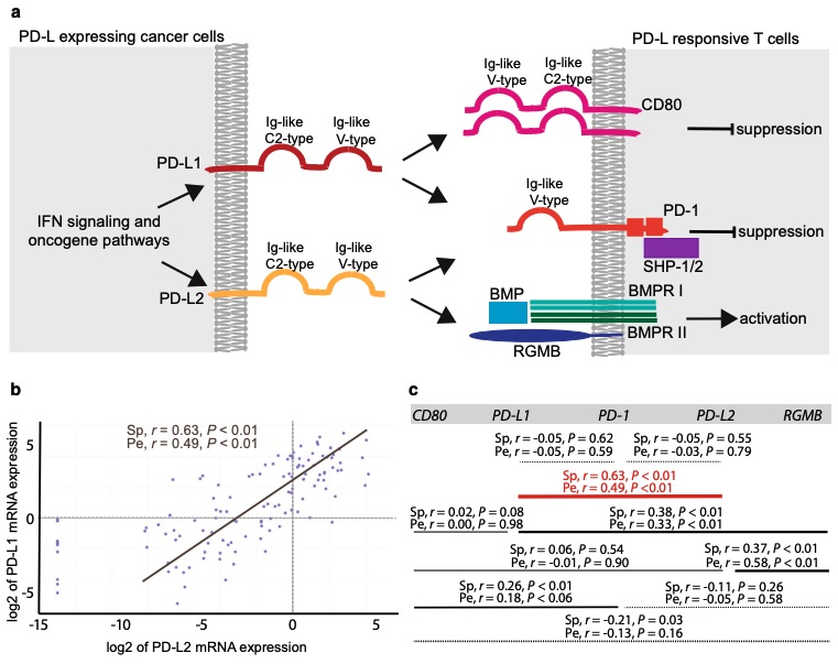 Fig. 1 PD-L1 and PD-L2 expression correlation analyses in 114 NSCLC cell lines from the CCLE dataset. 1
hPD-1/hPD-L1/hPD-L2 Signal Pathway
The hPD-1 cytoplasmic tail contains an immunoreceptor tyrosine-based inhibitor motif (ITIM) and immunoreceptor tyrosine-based switch motif (ITSM), which can recruit tyrosine phosphatases (SHP-1 and SHP-2) to block T cell signal transduction. After hPD-1 binds to its ligands, it can inhibit the proliferation of T cells and the production of cytokines such as IL-2 and IFN-γ. The interactions of hPD-1 with its ligands can also mediate the function of B cells, specifically inhibiting the proliferation, differentiation, and Ig secretion of B cells. hPD-1 interacts with hPD-L1 and hPD-L2 could result in the apoptosis of antigen-specific T cells in lymph nodes, while restraining the apoptosis of regulatory T cell (Tregs). In tumor microenvironment influenced by proinflammatory cytokines (such as IL-4 and IFN-γ), the hPD-L1 expression on tumor cells can be up-regulated. And then PD-L1 interacts with hPD-1 which presents on the surface of tumor-specific CD8+ T cells, to limit the host's immune response.
Development of hPD-1/hPD-L1/hPD-L2 Triple Humanized Mice
hPD-1 on T cells can be combined with both hPD-L1 on tumor cells and hPD-L2 on immune cells which infiltrate in the tumor to make T cells incompetent. Blockade the single hPD-1/hPD-L1 pathway might not be enough to activate the immunity to fight against the tumor cells. The triple combination of anti-hPD-1 antibody, anti-hPD-L1 antibody, and anti-hPD-L2 antibody is considered as a powerful strategy in cancer treatment. Creative Biolabs has advanced the drug development for our global clients using our well-established Hi-Affi™ “humanized” animal models. With years of CRO services, we have accumulated much experience in preclinical novel drug development. If you have any problems in your research, please feel free to contact us for further discussions and our scientists will provide you with scientific solutions to assist you in achieving your study goals.
Creative Biolabs also offers other various Humanized Mouse Models you may be interested in:
Reference
- Larsen, Trine Vilsbøll, Dianna Hussmann, and Anders Lade Nielsen. "PD-L1 and PD-L2 expression correlated genes in non-small-cell lung cancer." Cancer Communications 39 (2019): 1-14. Distributed under Open Access license CC BY 4.0. The image was modified by extracting and using a-c parts of the original image.
For Research Use Only.