Pulmonary Hypertension induced Right Heart Failure Modeling & Pharmacodynamics Service
Creative Biolabs proudly offers a diverse portfolio of meticulously characterized and well-established HF models, enabling comprehensive evaluation of novel drug candidates and therapeutic strategies.
Introduction
Heart failure (HF) represents a complex and debilitating syndrome where the heart struggles to pump sufficient blood to meet the body's metabolic demands. It is a leading cause of hospitalization and mortality worldwide, presenting a significant global health challenge. Developing effective therapeutic interventions necessitates robust and physiologically relevant preclinical models.
Pulmonary Hypertension-Induced Right HF Model
Pulmonary hypertension (PH) imposes a severe and sustained burden on the right ventricle (RV), ultimately leading to its hypertrophy, dysfunction, and eventual failure. Creative Biolabs offers highly translational rodent models to investigate this critical disease progression and evaluate therapeutic strategies.
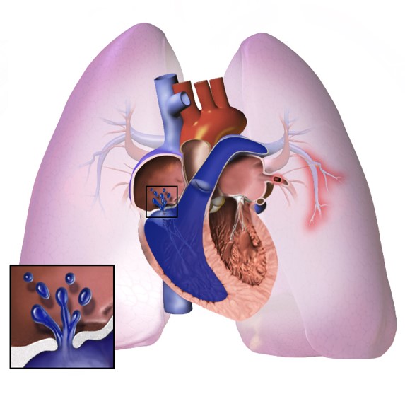 Fig.1 Schematic of pulmonary hypertension.Distributed under CC BY-SA 4.0, from Wiki, without modification.
Model Construction Steps
Our primary rodent models for PH-induced right HF (RHF) include the monocrotaline (MCT)-induced PH-RHF model and the chronic hypoxia-induced PH-RHF model.
MCT-Induced Model
01Preparation
Healthy rats or mice are acclimatized and weighed.
02Induction
A single subcutaneous injection of MCT is administered at a precisely calculated dose.
03Progression
Animals are monitored for a period of 3 to 4 weeks, during which the MCT induces pulmonary vascular endothelial damage, leading to pulmonary arterial remodeling and progressive PH, culminating in RV hypertrophy and dysfunction.
04Intervention
Therapeutic interventions typically commence after disease establishment, allowing for assessment of their impact on both PH and RHF progression.
Chronic Hypoxia-Induced Model
01Preparation
Rodents are placed in specialized hypoxic chambers.
02Induction
Animals are continuously exposed to a low-oxygen environment (e.g., 10% O2) for 3 to 6 weeks.
03Progression
The sustained hypoxia triggers pulmonary vasoconstriction and vascular remodeling, leading to PH and subsequent RV hypertrophy.
04Intervention
Therapeutic agents can be administered throughout or after the hypoxic exposure to assess their efficacy.
Strengths and Limitations
Strengths:
- Reproducibility: These models offer high reproducibility, ensuring consistent results across experiments.
- Well-Characterized: Extensive historical data support their utility, providing clear benchmarks for disease progression.
- Cost-Effective: Rodent models are generally more economical, facilitating larger-scale studies and screening.
- Mechanistic Insights: They allow for detailed investigation into specific cellular and molecular pathways underlying PH-RHF.
Limitations:
- Translational Gaps: Rodent physiology differs from humans, potentially limiting direct translatability of some findings.
- Acute vs. Chronic: The MCT model, while progressive, can be more acute than the chronic nature of human PH.
- Species-Specific Responses: Certain drug effects or disease mechanisms may vary between rodent species and humans.
Evaluation Platform
Creative Biolabs utilizes a comprehensive platform of advanced instruments and assays to assess disease progression and therapeutic efficacy. Our evaluation encompasses biochemical, molecular, cellular, histopathological, behavioral, and imaging modalities. Key indicators include:
- Invasive Hemodynamic Measurements: Gold-standard assessments of Right Ventricular Systolic Pressure (RVSP), Pulmonary Arterial Pressure (PAP), and Cardiac Output via Pressure-Volume (PV) Loops.
- Echocardiography: High-resolution non-invasive imaging for functional and structural insights, including RV dimensions, ejection fraction (EF), and Tricuspid Annular Plane Systolic Excursion (TAPSE).
- Histopathological Analysis: Thorough examination of tissue samples for Right Ventricular Hypertrophy Index, myocardial fibrosis, myocyte hypertrophy, and inflammatory cell infiltration.
- Gene and Protein Expression Profiling: Molecular techniques to analyze gene (e.g., qPCR, RNA-Seq) and protein (e.g., Western blot, proteomics) alterations in lung and heart tissues, providing mechanistic insights.
- Behavioral Assessment: Functional parameters like exercise capacity to gauge disease severity and treatment response.
- Survival Analysis: Monitoring survival rates as a critical endpoint reflecting therapeutic efficacy.
Applications
- Comprehensive Disease Simulation: Our models accurately simulate various forms of PH, including idiopathic, connective tissue disease-associated, congenital, and drug- or toxin-induced PH, providing relevant preclinical platforms for disease understanding.
- Diverse Therapeutic Evaluation: They enable the rigorous evaluation of numerous drug classes, such as vasodilators, anti-proliferative, anti-fibrotic, metabolic modulators, and RV-specific agents, assessing their impact on both pulmonary vascular and right ventricular function.
- Broad Modality Testing: The platform is suitable for testing a wide array of therapeutic modalities, from small molecule compounds and biologics (e.g., antibodies, peptides) to advanced gene, RNA-based, and cellular therapies aimed at restoring cardiovascular health.
Related Heart Failure Models
PA Constriction induced Right HF Model
Ascending Aortic Arch Constriction induced Post-Pressure Overload Heart Failure Model
Abdominal Aortic Stenosis induced Left HF Model
DOCA & Salt induced Left HF Model
Our Advantages
- Expert Scientific Team: Years of specialized experience in cardiovascular and respiratory preclinical research.
- Validated Models: Access to meticulously developed and consistently reproducible PH-RHF models.
- Comprehensive Readouts: State-of-the-art facilities for in-depth hemodynamic, imaging, and molecular analysis.
- Customizable Study Designs: Flexible experimental protocols tailored precisely to your specific research objectives.
- Translational Impact: Generating high-quality, regulatory-compliant data to accelerate your drug development pipeline.
Work with Us
- Summarize the project requirements and fill in the information collection form.
- Sign a CDA from both parties to further communicate information, such as targets.
- Select an animal model, discuss experimental design, and determine assay parameters.
- Project costing and project schedule forecasting.
- We provide a detailed project plan, including the required sample quantities, methods, and protocols.
- Both parties confirm the project details and start the project.
- Confirm the timeline of the project.
- We provide periodic results and information on the animal's condition.
- We will work together to make project adjustments as necessary.
- We provide a comprehensive project report promptly.
- We arrange transportation for the produced samples.
- We provide a discussion of the project results and help to arrange the next steps.
- Data storage and archiving.
Contact Us
Creative Biolabs is dedicated to supporting your research endeavors with our specialized PH-Induced RHF Model services. Our team provides expert guidance and customized solutions from initial study design through final data interpretation. We encourage you to reach out to us to discuss how our robust preclinical platforms can advance your cardiovascular drug discovery program.
FAQs
-
Q1: How is right ventricular function accurately measured in these models?
A: Right ventricular function is precisely assessed using a combination of invasive and non-invasive methods. We perform gold-standard right heart catheterization to obtain hemodynamic parameters like RV systolic pressure and pulmonary arterial pressure, complemented by pressure-volume loop analysis for comprehensive contractility and relaxation metrics. Non-invasively, high-resolution echocardiography provides detailed functional and structural insights, including RV dimensions, ejection fraction, and tricuspid annular plane systolic excursion (TAPSE).
-
Q2: What histological endpoints are critical for assessing right ventricular remodeling?
A: For assessing right ventricular remodeling, critical histological endpoints include calculating the right ventricular hypertrophy index (ratio of RV weight to left ventricle plus septum weight), quantifying collagen deposition to evaluate fibrosis, measuring myocyte cross-sectional area to gauge hypertrophy, and identifying the presence and extent of inflammatory cell infiltration within the myocardial tissue.
-
Q3: Can your models differentiate between primary PH effects and those specifically impacting the RV?
A: Indeed, certain models within our platform are designed to delineate these effects. For instance, surgical models like pulmonary artery banding (PAB), when adapted for rodents, primarily induce a pressure overload on the RV. This allows researchers to focus specifically on the adaptive and maladaptive responses of the RV independently of the primary pulmonary vascular disease.
-
Q4: How do you ensure the reproducibility and consistency of your PH-RHF models?
A: Our commitment to high reproducibility stems from strict adherence to meticulously standardized operating procedures for animal husbandry, disease induction, and all subsequent measurements. We implement rigorous quality control checks at every stage and rely on our team of highly trained and experienced technical staff to ensure consistent execution across all experimental cohorts.
-
Q5: Are your models suitable for evaluating the long-term effects of chronic therapeutic interventions?
A: Yes, our flexible study designs are well-suited for assessing the long-term impact of chronic therapeutic interventions. We can accommodate extended dosing regimens over prolonged periods, allowing for a thorough evaluation of sustained therapeutic efficacy and any potential long-term adverse effects on both the pulmonary vasculature and the critical function of the right heart.
Published Data
1. MCT-Induced Model
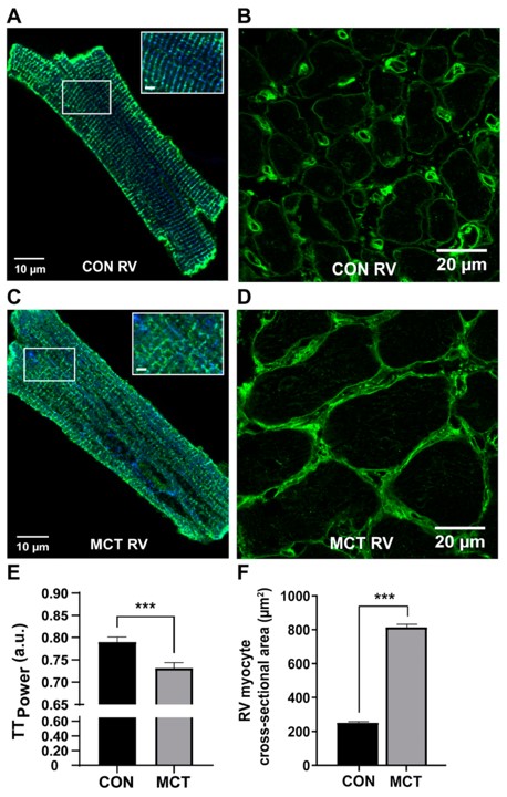 Fig.2 Myocardial Changes in Response to MCT.1
A recent review highlighted that the MCT rat model provides a consistent and inexpensive means to study the progression from compensated right ventricular (RV) hypertrophy to RV failure induced by PH. Project results from studies using this model have shown clear evidence of RV hypertrophy, increased RV thickness, and cardiomyocyte enlargement at 4 weeks post-MCT injection, along with attenuated body weight, mirroring key aspects of human disease progression. This underscores the model's utility for investigating underlying cellular mechanisms and evaluating novel therapeutic targets.
2. Chronic Hypoxia-Induced Model
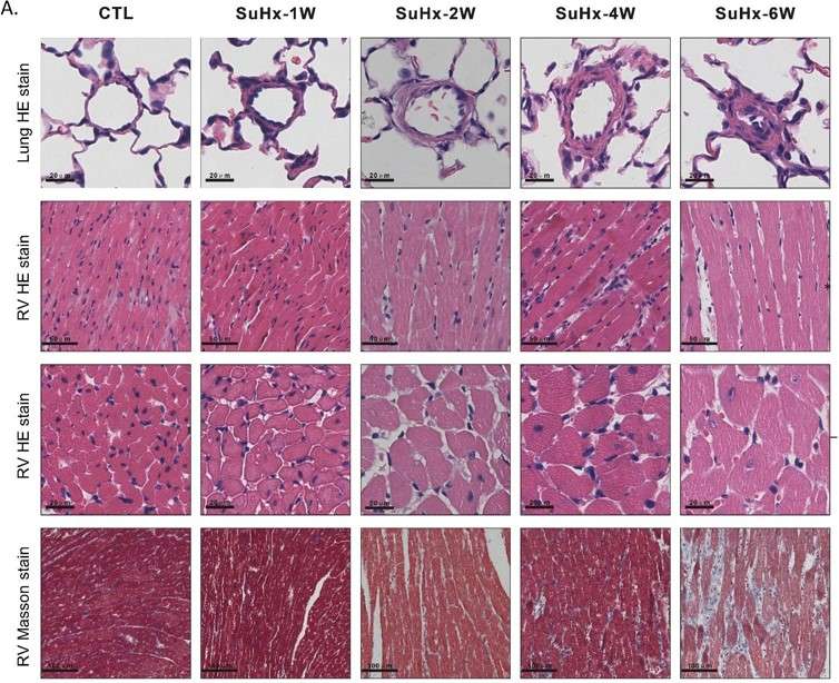 Fig.3 Changes of pulmonary vascular medial wall thickness and RV lesions.2
A study demonstrated the intricate relationship between DNA damage response and RHF progression in a SU5416-hypoxia-induced PH rat model. Project results showed that right ventricular pressure and hypofunction manifested earlier than pulmonary artery thickening. This indicated that right ventricular structural and functional deterioration, linked to DNA damage and cellular senescence, begins in the early stages of PH development. This underscores the model's relevance in understanding disease progression and identifying early intervention targets.
References
- Krstic, Anna Maria et al. "The Monocrotaline Rat Model of Right Heart Disease Induced by Pulmonary Artery Hypertension." Biomedicines vol. 12,9 1944. 23 Aug. 2024. Distributed under Open Access license CC BY 4.0, without modification. https://doi.org/10.3390/biomedicines12091944
- Kuang, Meidan et al. "Echocardiographic evaluation of right heart failure which might be associated with DNA damage response in SU5416-hypoxia induced pulmonary hypertension rat model." Respiratory research vol. 24,1 202. 17 Aug. 2023. Distributed under Open Access license CC BY 4.0, without modification. The image was modified by extracting and using only part of the original image. https://doi.org/10.1186/s12931-023-02501-7
For Research Use Only.