Rheumatoid Arthritis (RA) Modeling & Pharmacodynamics Services
Creative Biolabs offers a range of well-established and customizable models to assess the efficacy of RA therapeutics. These models simulate the pathophysiology of RA, including joint inflammation and immune cell infiltration, providing a reliable platform for evaluating novel drug candidates and therapeutic interventions.
Introduction
Rheumatoid arthritis (RA) is a chronic autoimmune disease characterized by inflammation of the synovial joints, leading to pain, swelling, stiffness, and eventually joint destruction. It affects approximately 1% of the global population, with a higher prevalence in women. RA occurs when the immune system mistakenly attacks the body's own tissues, particularly the joints, causing inflammation that can spread to other organs, including the skin, lungs, and heart. Over time, unchecked inflammation leads to irreversible damage to the joint structures, including cartilage, bone, and ligaments, resulting in disability and decreased quality of life. RA is a multifactorial disease with genetic, environmental, and immunological factors contributing to its onset and progression. The treatment landscape for RA includes nonsteroidal anti-inflammatory drugs (NSAIDs), disease-modifying antirheumatic drugs (DMARDs), and biologic agents that target specific immune pathways. However, there remains a need for more effective and targeted therapies that can address the underlying causes of RA and prevent joint damage.
RA Models and Applications
Creative Biolabs offers a comprehensive range of well-established rodent models for studying rheumatoid arthritis (RA). These models are carefully designed to replicate key aspects of human RA, such as joint inflammation, cartilage destruction, and systemic immune response. They are accompanied by thorough evaluations of various clinical and pathological parameters, enabling precise assessment of therapeutic candidates during the preclinical phase. Our team of experienced scientists will support you throughout your project, from experimental design to data interpretation, ensuring reliable and high-quality results. To learn more about the rheumatoid arthritis models available for preclinical research, please explore the links below:
| Model | Simulates Disease | Evaluates Drugs | Animal species |
| Adjuvant induced Arthritis (AIA) Model | Simulates acute inflammation and chronic joint damage, mimicking early stages of rheumatoid arthritis. | Anti-inflammatory drugs, corticosteroids, immunosuppressive agents, and biologics (e.g., TNF inhibitors). | Rat, Mouse |
| Collagen induced Arthritis Rodent Model | Simulates the chronic autoimmune arthritis seen in RA, including autoantibody production and joint inflammation. | Disease-modifying antirheumatic drugs (DMARDs), biologics targeting TNF, IL-6, and B cells. | Rat, Mouse |
| Collagen Antibody induced Arthritis (CAIA) Model | Induces joint inflammation via autoantibody administration, closely mimicking late-stage RA in humans. | Biologics targeting specific immune responses, small molecule inhibitors, and novel immunotherapies. | Rat, Mouse |
| Antigen induced Arthritis Model | Induces arthritis through immune response to specific antigens, resembling certain aspects of RA pathophysiology. | Anti-inflammatory agents, cytokine blockers, immunomodulatory drugs, and biologics targeting the immune system. | Rat, Mouse |
| Zymosan A (ZIA) induced Rheumatoid Arthritis (RA) Model | Induces acute inflammation in joints via immune activation, often used for evaluating inflammation-driven drug efficacy. | Anti-inflammatory drugs, pain management therapies, and agents targeting inflammatory mediators like IL-1β. | Mouse |
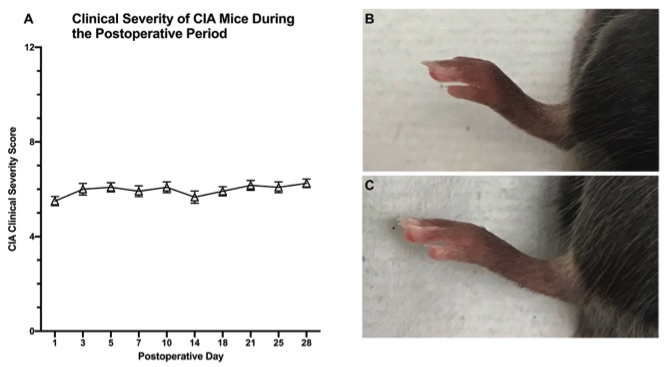 Fig. 1 Experimental results Of mice induced with CIA.1,3
Evaluation Platform
- Animals: Mouse, Rat, NHPs.
-
Measurements
We offer a variety of measurements for evaluating drug efficacy in rheumatoid arthritis models, utilizing an array of advanced technologies, including but not limited to:- General observations: Joint swelling, deformity, body weight, and mobility.
- Histopathological analysis: Tissue samples are examined for synovial inflammation, cartilage destruction, and bone erosion through H&E staining and Masson's trichrome staining.
- Cytokine profiling: Measuring inflammatory cytokines such as TNF-α, IL-1β, IL-6, and IL-17 via ELISA, qPCR, or Western blotting.
- Immunohistochemistry: Assessing immune cell infiltration, such as T-cells and macrophages, in synovial tissue.
- Radiographic analysis: X-ray imaging to evaluate joint destruction and bone erosion.
- Serum biomarkers: Measuring serum levels of inflammatory markers like CRP (C-reactive protein) and rheumatoid factor (RF).
- Gene/protein expression profiling: Using RT-PCR and Western blot to evaluate key inflammatory pathways involved in RA progression.
Our advantages
- Comprehensive Drug Testing: Our models allow for in-depth testing of a wide range of therapeutic approaches, from conventional treatments to biologic therapies.
- High Reproducibility: The models are highly reproducible, ensuring reliable and consistent results across experiments.
- Customized Solutions: We offer flexible models tailored to specific research needs, whether for acute or chronic disease phases.
- Expert Guidance: Our experienced scientific team provides expert assistance in study design, protocol optimization, and data analysis, ensuring optimal experimental outcomes.
- Multi-Platform Integration: We integrate various measurement platforms like histology, radiography, and cytokine profiling to provide a comprehensive assessment of drug efficacy.
Work with Us
- Summarize the project requirements and fill in the information collection form.
- Sign a CDA from both parties to further communicate information, such as targets.
- Select an animal model, discuss experimental design, and determine assay parameters.
- Project costing and project schedule forecasting.
- We provide a detailed project plan, including the required sample quantities, methods, and protocols.
- Both parties confirm the project details and start the project.
- Confirm the timeline of the project.
- We provide periodic results and information on the animal's condition.
- We will work together to make project adjustments as necessary.
- We provide a comprehensive project report promptly.
- We arrange transportation for the produced samples.
- We provide a discussion of the project results and help to arrange the next steps.
- Data storage and archiving.
FAQs
-
1. What are the most commonly used methods to induce RA in animal models?
The most common methods are collagen induced arthritis (CIA), complete Freund's adjuvant (CFA) injection, and the K/BxN serum transfer model. These methods trigger an immune response that leads to joint inflammation.
-
2. Can this model be used for evaluating both acute and chronic RA treatments?
Yes, this model can simulate both acute inflammation and chronic joint damage, making it ideal for evaluating treatments at different stages of disease progression.
-
3. Are the RA models customizable to study specific aspects of the disease?
Absolutely. We offer customizable models that focus on specific aspects of RA, such as immune cell dynamics, joint destruction, or the systemic effects of the disease.
-
4. How long does it take to establish an RA model?
Typically, the disease model can be established within 2-3 weeks following induction, with chronic inflammation developing over several weeks for long-term studies.
-
5. What are the advantages of using this model for RA drug testing?
This model mimics key features of human RA, such as joint inflammation, immune cell infiltration, and cartilage destruction, making it ideal for screening RA therapeutics.
Published Data
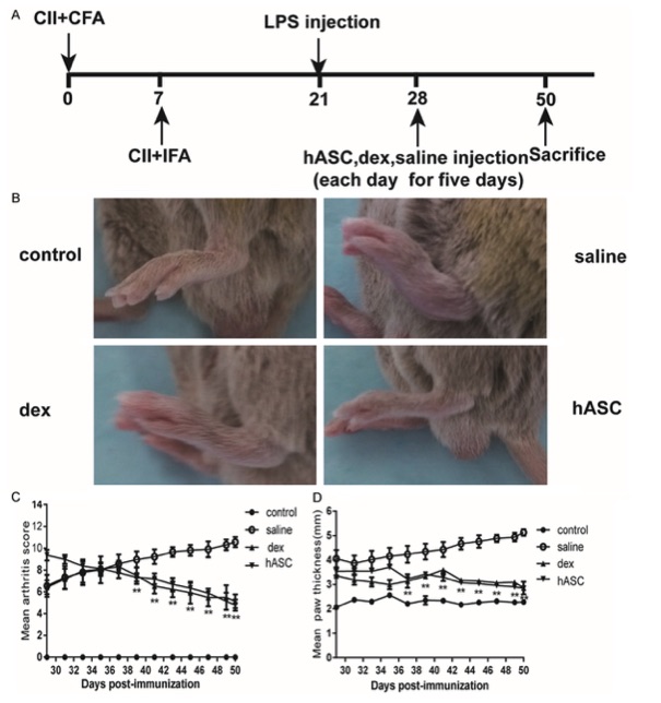 Fig. 2 Therapeutic effects of hASCs in CIA mice.2,3
Collagen induced arthritis (CIA) was induced in DBA/1 mice by injecting them with bovine type II collagen (CII) emulsified in complete Freund's adjuvant (CFA), followed by a booster injection of CII emulsified in incomplete Freund's adjuvant (IFA) seven days later. On day 21, the mice received an injection of lipopolysaccharide (LPS). From day 28, daily intravenous injections of adipose-derived stem cells (ASCs), dexamethasone (dex), or saline were administered (Figure 2A). Arthritis development was monitored up to day 50, with daily evaluations of arthritis severity. Photographs of the paws on day 50 post-induction are presented in Figure 2B, showing that hASC treatment significantly reduced edema and erythema in the arthritic joints compared to the CIA group. As demonstrated in Figures 2C and 2D, hASC administration in mice with established arthritis (arthritis score >2) led to a marked reduction in disease severity and hind paw thickness compared to saline-treated controls.
References
- Trikha, Rishi et al. "Active rheumatoid arthritis in a mouse model is not an independent risk factor for periprosthetic joint infection." PloS one vol. 16,8 e0250910. 16 Aug. 2021. https://doi.org/10.1371/journal.pone.0250910
- Zhang, Li et al. "Use of immune modulation by human adipose-derived mesenchymal stem cells to treat experimental arthritis in mice." American Journal of Translational Research vol. 9,5 2595-2607. 15 May. 2017
- Distributed under Open Access license CC BY 4.0, without modification.
For Research Use Only.