Cell Scratch Assay
Creative Biolabs focuses on cell scratch assay to meet the needs of customers for scientific research and related biomedical industries.
Cell Scratch Assay to Quantity the Migration of Tumor Cell
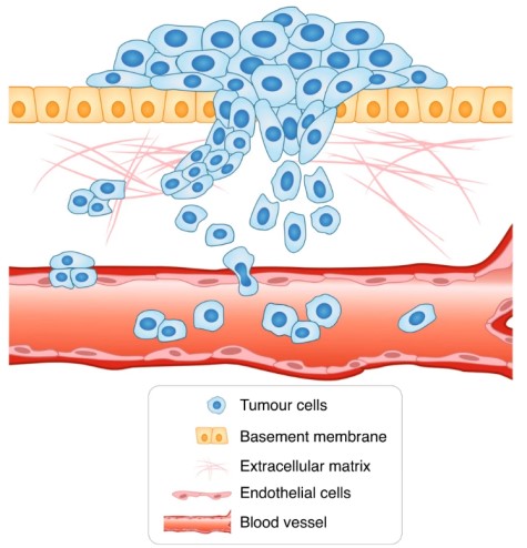
Cell migration is a hallmark of tumor development and is often associated with angiogenesis to ensure tumor nutrition, as well as metastasis formation. Developing methods to study migration is essential to study the mechanisms of tumorigenesis.
Cell scratch assay is a commonly used assay to test cell migration as it is economical and easy to perform on adherent cell lines such as endothelial cells, epithelial cells, and fibroblasts. Creative Biolabs offers in vitro cell scratch assay services to assist customers' projects in cancer research.
 Fig.1 The model of cancer cell migration and invasion. (Novikov, 2021)
Assay Procedure
In cell scratch experiments, an adherent cell line that binds properly to the bottom of the well is generally required to detect and analyze migration. In order to measure migration, we usually modify the appropriate culture conditions in advance, such as reducing the serum concentration, starving the cells before scraping or using some inhibitors. Root according to the different cell types to choose the appropriate measures. After pre-culturing adherent cells for a period of time, uniform scratches will be carried out mechanically with pins in 96-well format. After the application of the test compound, the migration of incubated cells into the scratched area is detected by the instrument in real-time.
 Fig.2 The scheme of the cell scratch assay process. (Creative Biolabs)
Attractive Advantages
- Well-established and Easy Assay
- In Vitro Cell Migration Detection
- Real-time Monitoring
- No Labeling Required
- Expert research team
Case Study
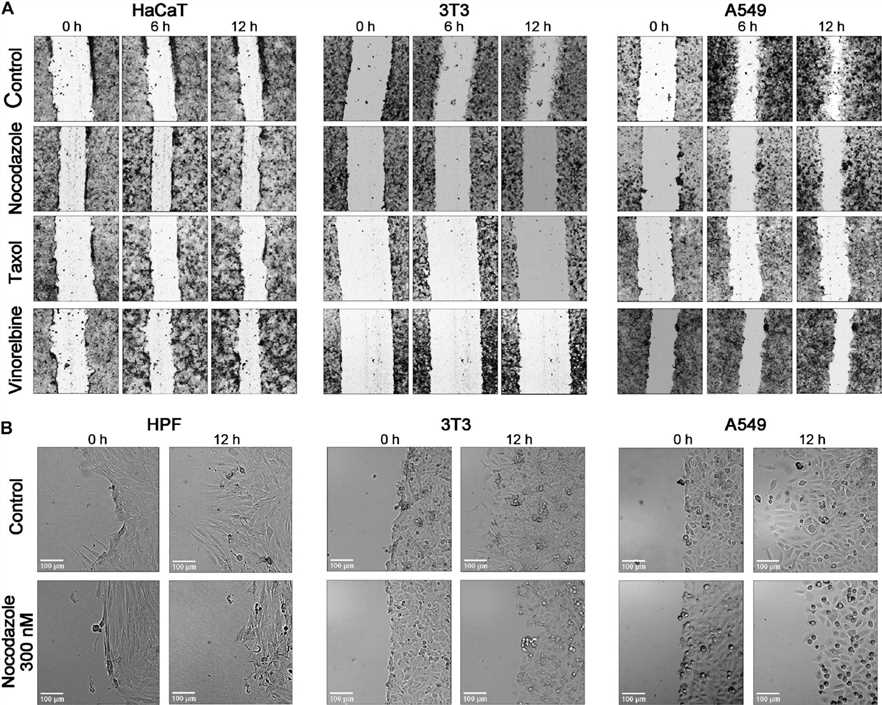
| Background | Cell migration is a fundamental process in various physiological events like wound repair and tumor metastasis, heavily reliant on the actin cytoskeleton and microtubules. Microtubule-interfering drugs can impede cell migration, but how they do this is not fully understood. The wound healing assay is a popular method to study cell motility, but it often yields semi-quantitative results due to limitations in image analysis. There's a need for more precise tools to analyze cell migration and the effects of drugs on it. |
| Method | The researchers developed an automated pipeline to segment and analyze images from wound healing assays. This pipeline was tested for accuracy and used to study the effects of microtubule inhibitors on various cell lines. The experiments involved live-cell imaging with frequent sampling (10-minute intervals) over 24 hours, and the analysis focused on a specific 6-hour window of linear wound closure. |
| Result | The study found that wound closure dynamics are often non-linear, but a linear phase exists between the 5th and 12th hours post-scratch. Within this linear window, the researchers quantified the effects of microtubule inhibitors on cell motility. The inhibitors generally slowed down cell motility at higher concentrations, but the response varied across cell lines and drugs. Interestingly, low doses of these drugs sometimes increased cell motility. |
 Fig.2 Comparing the temporal course of wound closure with anti-MT medications.2
Customer Speaking
Ju***a 17/May/22 ⭐⭐⭐⭐⭐
For a sequence of cell movement investigations connected to a cancer research project, our lab required a consistent and fast turnaround. The cell scratch test program offered by Creative Biolabs completely matched what we were seeking. The procedure was simple, hence it was fantastic that we didn't have to spend much time labeling cells. Another great advantage was the real-time monitoring, which produced rather comprehensive data. Our findings were within the expected range, and the data analysis they supplied was straightforward and understandable.
Frequently Asked Questions
Q: Why choose cell scratch assay?
A: The cell scratch assay is a low-cost, easy, and well-developed experiment type to detect cell migration in vitro. In addition, the assay enables real-time monitoring of cell migration and does not require labeling.
Q: Which cell types does this assay apply to?
A: Adherent cell lines such as endothelial cells, epithelial cells, and fibroblasts.
For more details about our cell scratch assay services, please don't hesitate to contact us for more information.
References
- Novikov, Nikita M., et al. "Mutational drivers of cancer cell migration and invasion." British Journal of Cancer 124.1 (2021): 102-114. Distributed under Open Access License CC BY 4.0, without modification.
- Kauanova, Sholpan, Arshat Urazbayev, and Ivan Vorobjev. "The frequent sampling of wound scratch assay reveals the “opportunity” window for quantitative evaluation of cell motility-impeding drugs." Frontiers in Cell and Developmental Biology 9 (2021): 640972. Distributed under Open Access License CC BY 4.0, without modification.
For Research Use Only.
