Endothelin-1 induced Stroke & Brain Ischemia Modeling & Pharmacodynamics Service
Creative Biolabs has accumulated profound expertise in conducting stoke preclinical studies. Our comprehensive battery of ischemic stroke models involves focal ischemia and global ischemia, transient and permanent occlusions. moreover, we provide stoke models for both rat and mouse species. Especially, we utilize a focal ischemic stroke model, endothelin-1 model, for the evaluating of candidate pro-regenerative therapies and for the screening of drugs developed for the treatment of thromboembolic stroke.
Induction of Endothelin-1 Model
Endothelin-1 (ET-1) is a potent and long-acting vasoconstrictive peptide which is produced endogenously during ischemic stroke and which contributes to overall loss of cells and disability. As a result, this model of focal stroke is based on the application of exogenous ET-1, which induces stroke and cell death after sustained vasoconstriction with reperfusion. It can be microinjected to induce a focal stroke in small tissue volumes (e.g., cortical grey matter, white matter or subcortical tissue) or after injection near the middle cerebral artery (MCA). This model can be induced in both rats and mice, but rats are more frequently used. Laser Doppler flowmetry (LDF) can be used to verify cerebral ischemia during ET-1 infusion.
Features of Endothelin-1 Model
- The procedure is simple and can be performed quickly.
- It can be used in elderly rats with only very low resulting mortality.
- The artery constriction can be controlled by altering the dose of ET-1 delivered.
- There is no need to manipulate the extracranial vessels supplying blood to the brain.
- It shows gradual reperfusion rates that more closely mimic the reperfusion in humans.
- It has the ability to avoid anesthesia by infusion of ET-1 in conscious rats.
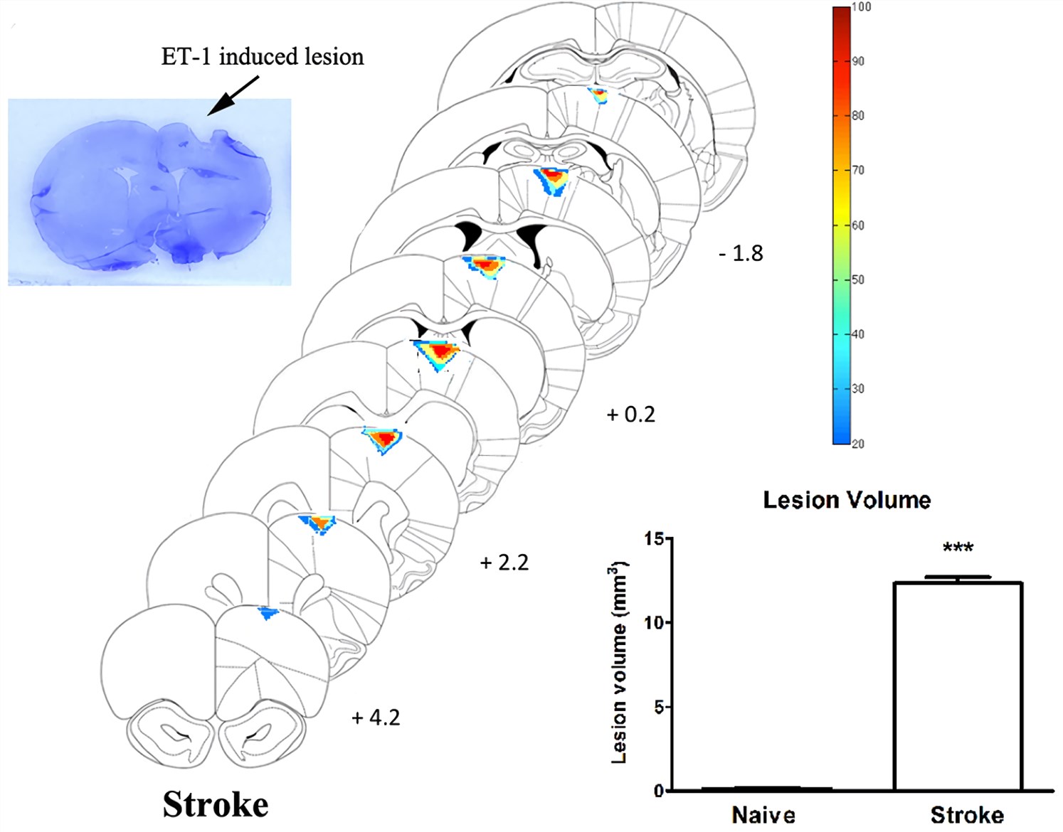 Fig.1 Histological examination of stroke lesion location and volume. (Chan et al. 2017)1, 2
Assessments
Our preclinical service section offers an array of behavioral tests, functional recovery test, and histological assessments to assess the potential effect of novel therapeutics. Tests that are used to evaluate neurological deficits include balance, grip strength, paw placing, postural asymmetry, and staircase climbing. Moreover, brain sections of test animals are stained with 2,3,5-triphenyltetrazolium chloride (TTC) to evaluate the infarct volume, or we can use the magnetic resonance imaging (MRI) technique. Creative Biolabs offers assessments including but not limited to:
- Modified neurological severity score
- Behavioral tests (e.g., motor function, cognition, social behavior)
- Infarct volume measurement
Creative Biolabs also offers other stroke and brain ischemia rodent models that you may be interested in:
- Middle Cerebral Artery Occlusion (MCAO) Model (transient/permanent)
- Photothrombotic Ischemia Model
- Global Ischemic Four-Vessel Occlusion (4-VO) Rat Model
The comprehensive list of rodent neurological disease models is placed below for your review. Please click the links for more detailed description of each model:
We provide guidance regarding which models (species, transient or permanent ischemic stroke models) would be the most appropriate for your technology. Additionally, Creative Biolabs' in vivo pharmacology specialists can work with you to custom a detailed study design to your exact specifications. Contact us to discuss your specific requirements.
References
- Chan, H. H.; et al. Crossed cerebellar atrophy of the lateral cerebellar nucleus in an endothelin-1-induced, rodent model of ischemic stroke[J]. Frontiers in Aging Neuroscience. 2017, 9:10.
- under Open Access license CC BY 4.0, without modification.
For Research Use Only.