CTL ELISPOT Assay
Background of CTL ELISPOT Assay
The evaluation of immune responses is essential for understanding the effectiveness of biotherapies and developing new treatment options. One important aspect of the immune response is cell-mediated cytotoxicity, which functions a lot in fighting pathogens and tumors. Creative Biolabs offers a CTL ELISPOT assay that can accurately measure cell toxicity T cell (CTL) activation and assess CTL immunity, providing valuable insights into the immune response, and helping researchers make informed decisions about the development of new treatments.
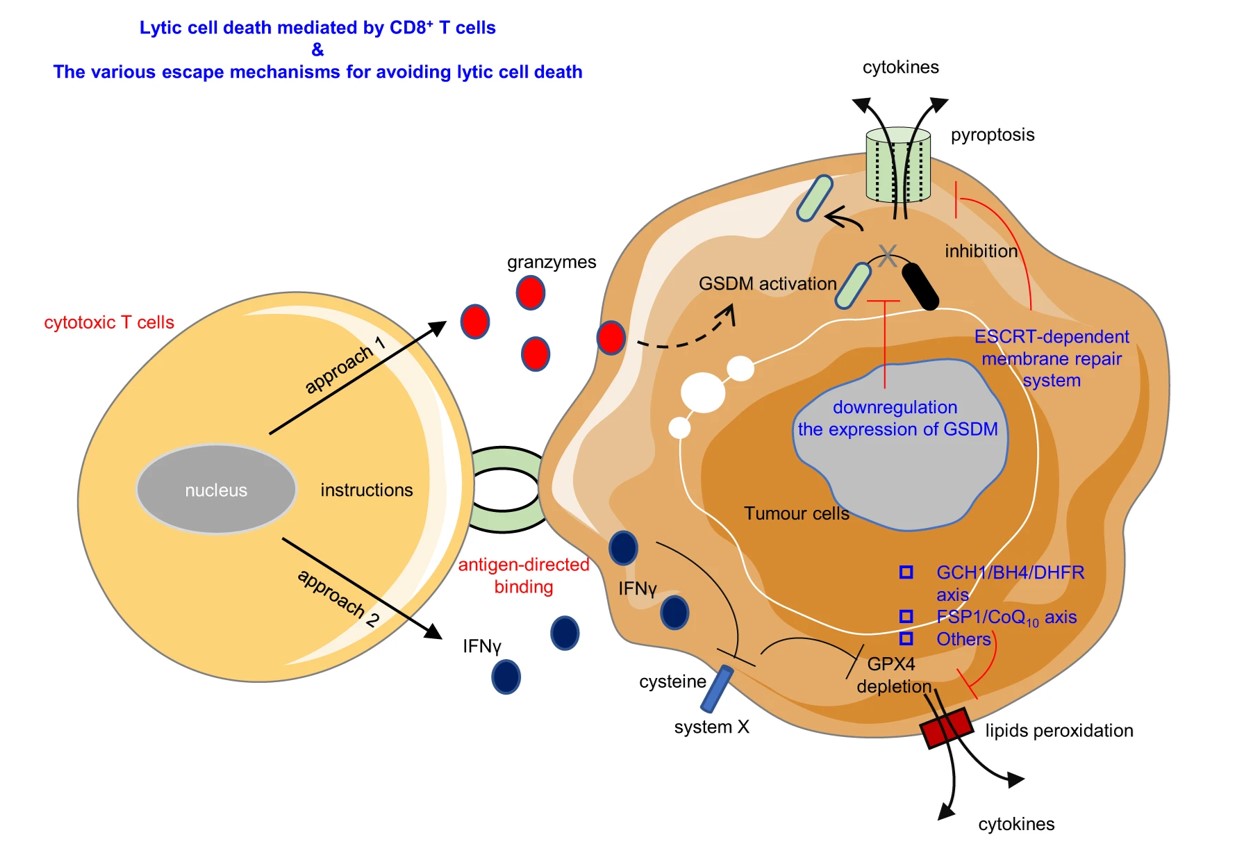 Fig.1 The cell lysis mediated by CD8+ T cells.1
CTL ELISPOT Assay at Creative Biolabs
Our CTL ELISPOT assay leverages advanced cell culture methods combined with enzyme-linked immunosorbent techniques to quantify cytokines secreted by individual CTL cells. This process employs coated antibodies to capture these cytokines from cultured CTL cells, followed by visualization through enzyme-linked spot color development. By directly stimulating live cells with specific antigens, we assess their functional responses in real-time.
Our assay is distinguished by its exceptional sensitivity, allowing for the detection of rare antigen-specific T cells that generate cytokines and effector molecules like perforin and granzyme B. Our assay bypasses the need for complex in vitro cell amplification, avoids the use of isotopes, and adheres to standardized protocols, allowing a single experimenter to efficiently handle hundreds of samples simultaneously. This greatly enhances throughput compared to traditional techniques. Importantly, from conceptual design to final result validation, our team of experts will provide comprehensive support, ensuring the successful completion of your project.
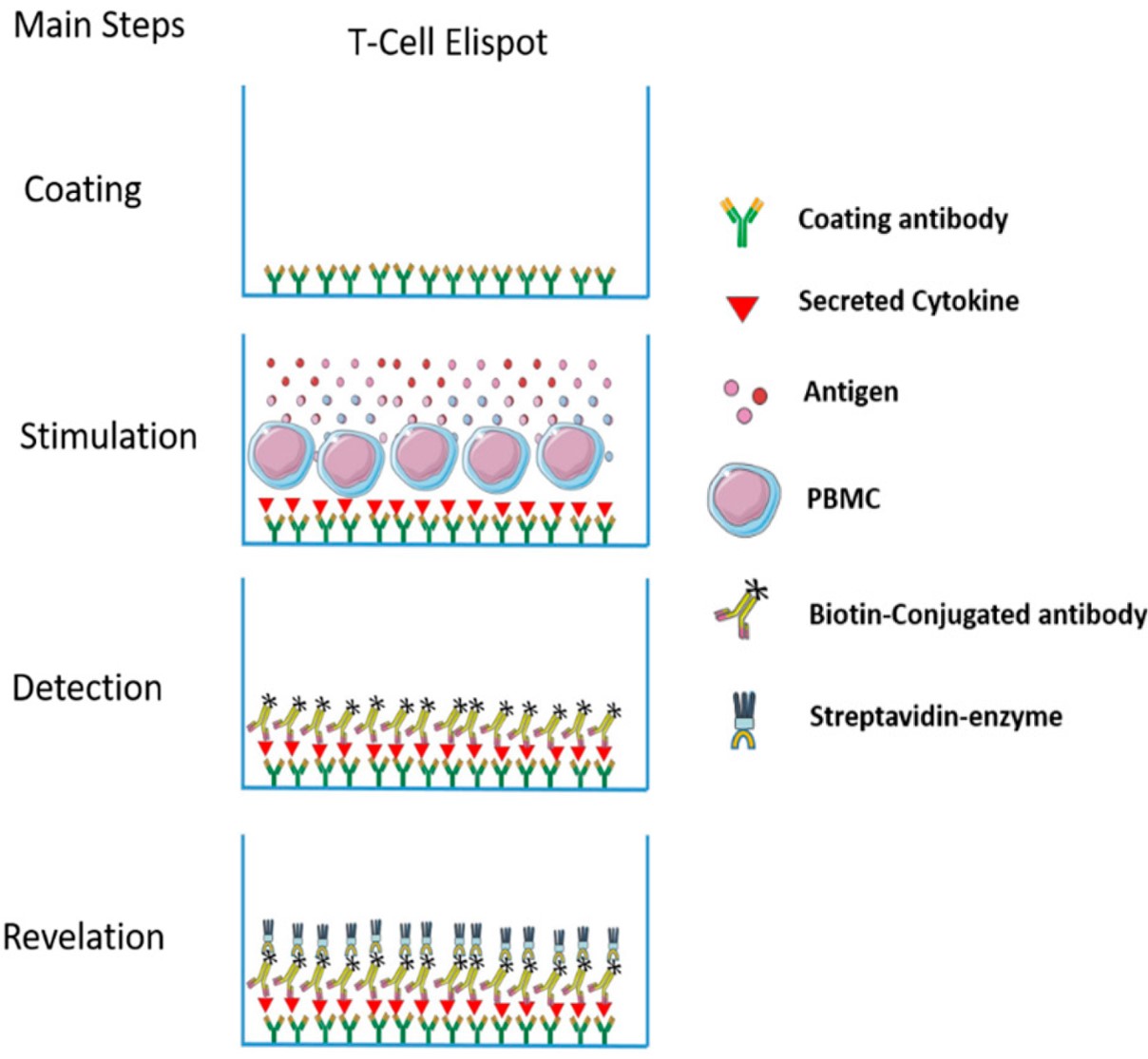 Fig.2 The main steps in CTL ELISPOT assay.2
Antigen Formats for CTL ELISPOT Assay
- Freeze and thaw lysate.
- Class I and class II peptides.
- Recombinant proteins.
- RNA.
- Recombinant virus vectors.
Our Advantages
- Exceptionally sensitive, capable of detecting cytokine secretion from as few as one cell among a million.
- Our assay allows direct stimulation with antigens and functional detection at the single live cell level.
- Easy and cost-effective, enabling high-throughput screening.
Data Show
This study focused on identifying SARS-CoV-2 peptide epitopes capable of binding to MHC class I molecules, with the goal of pinpointing viral protein-derived peptides that potentially trigger CD8+ T cell immune responses.
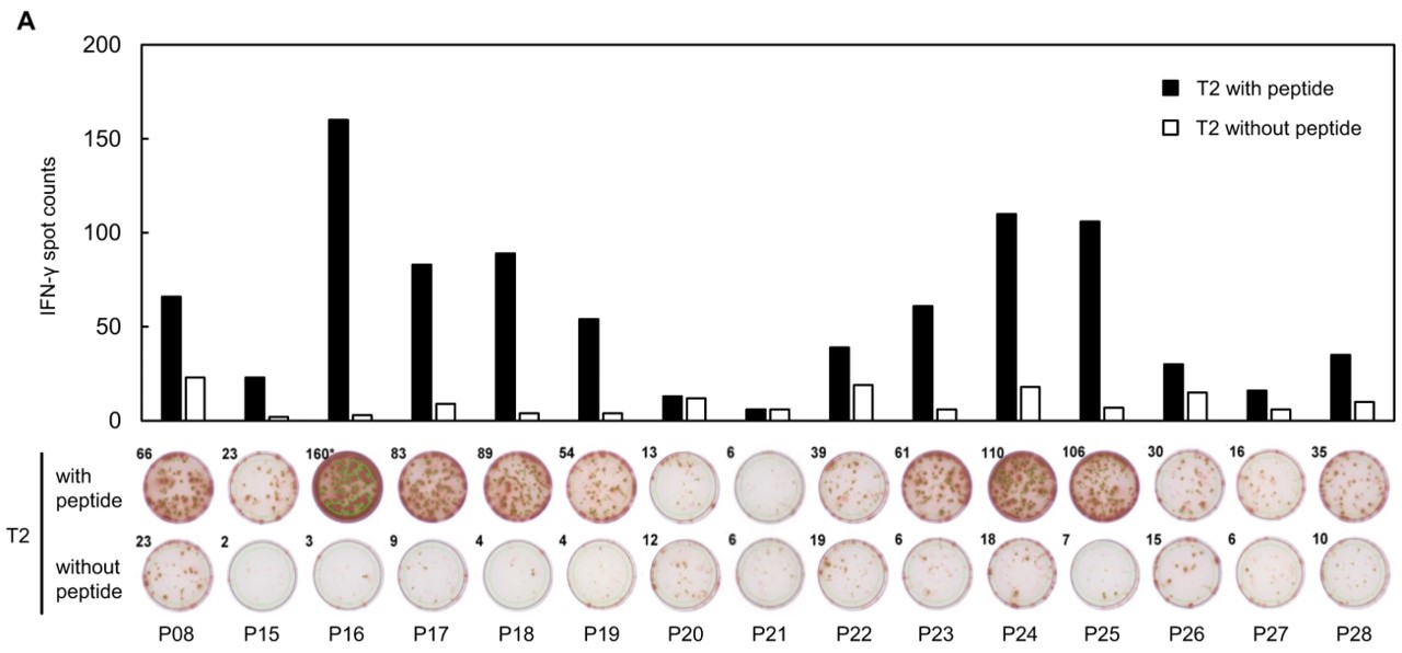 Fig.3 An IFN-γ ELISPOT assay was conducted following the in vitro induction of CTLs.3
Frequently Asked Questions
- Q1: What is the significance of small and large spots?
- Q2: What is the frequency range for precise measurements?
- Q3: What is the reason the spots are poorly defined or confluent in the CTL ELISPOT assay?
For more details about our CTL ELISPOT assay, please feel free to contact us.
References
-
Zuo, Zhigui, et al. "A cytotoxic T cell inspired oncolytic nanosystem promotes lytic cell death by lipid peroxidation and elicits antitumor immune responses." Nature Communications 14.1 (2023): 5456.
Distributed under Open Access License CC BY 4.0. The original image was modified by extracting and using part a, and the title was changed to "The cell lysis mediated by CD8+ T cells". -
Lima-Junior, Josue da Costa, Fernanda Nazaré Morgado, and Fátima Conceição-Silva. "How can Elispot add information to improve knowledge on tropical diseases?." Cells 6.4 (2017): 31.
Distributed under Open Access License CC BY 4.0. The original image was modified by extracting and using a part, and the title was changed to "The main steps in CTL ELISPOT assay". -
Hikichi, Tetsuro, et al. "Identification of cytotoxic T cells and their T cell receptor sequences targeting COVID-19 using MHC class I-binding peptides." Journal of Human Genetics 67.7 (2022): 411-419.
Distributed under Open Access License CC BY 4.0. The original image was modified by extracting and using part A, and the title was changed to "An IFN-γ ELISPOT assay was conducted following the in vitro induction of CTLs".
For Research Use Only.