Hi-Affi™ hPD-1/hTLR9 Dual Humanized Mouse Model
Anti-PD-1 antibody is a breakthrough for human cancer treatment but it is not almighty in clinical therapy. It has been evidenced that in some cases anti-PD-1 antibodies have a low response rate or could develop resistance after a period of treatment. However, clinically combinational use of anti-toll-like receptor 9 (TLR9) agonist antibody and anti-PD-1 antibody has a significant effect on patients with malignant melanoma. Creative Biolabs has successfully established an optimized Hi-Affi™ “humanized” animal platform to offer specialty manipulated hPD-1/hTLR9 dual humanized mice for our clients all over the world.
hPD-1/hTLR9 Molecule
There is a protein on the immune cell called hPD-1 (human programmed cell death protein 1). Tumor cells will produce an immunoglobulin-like molecule called hPD-L1 (human programmed cell death ligand 1). The interaction of hPD-1 and hPD-L1 could transmit a downregulatory signal which inhibits the activation of cytotoxic T cells.
Human TLR9 (hTLR9) was discovered initially from the innate immune system and is expressed in dendritic cells (DCs), B cells, and macrophages. It was subsequently found in the human tracheal mucosal epithelium, digestive tract, and pancreas. In recent years, studies have confirmed that hematoma cells and various solid tumor cells, including glioma, breast, prostate, liver, ovarian, and esophageal cancer cells also have hTLR9 expression. hTLR9 expression is positively correlated with tumor invasion, metastasis, and poor prognosis
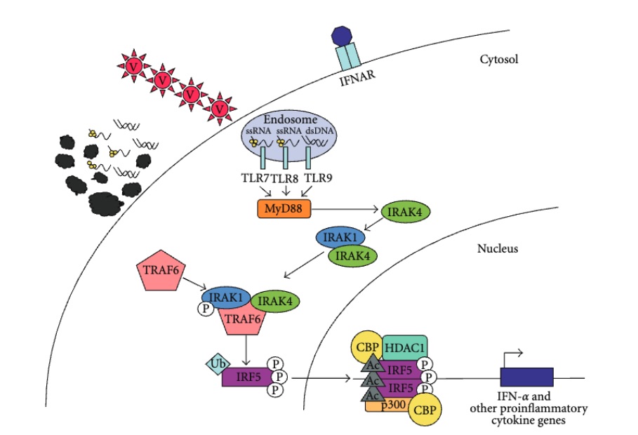 Fig. 1 Cells use TLRs as sensors to detect the presence of viruses (V) via TLR7, -8, and -9. 1
hPD-1/hTLR9 Signal Pathway
The binding of hPD-1 and hPD-L1 blocks the attack of cytotoxic T cells on tumor cells. Tumors adopt this method to hide themselves and survive. hTLR9 is initially localized in the cytoplasmic endoplasmic reticulum and transports to the lysosome via the membrane protein NC93B1. Here, it is combined with the ligand CpG DNA (synthesized as CpG ODN) which is endocytosed by the cell. The adapter molecule MyD88 is first recruited to form the hTLR9 complex (including MyD88, IRAK4, and adapter molecule TRAF6). IRAK4 phosphorylates IRAK1, activates IRAK, MAPK kinase, and interferon (IFN) regulators that interact with nuclear transcription factor κB (NF-κB), regulating gene expression and function. In malignant aggressive tumors, the expression level of the hTLR9 gene in tumor cell lines is always up-regulated. Specifically, hTLR9 causes the up-regulation of the transfer-related molecules such as IL-1/8, intercellular adhesion molecule-1, matrix metalloproteinase-2, and CXCR4, promotes the secretion of IL-10, and inhibits the production of anti-tumor cytokine IL-12.
Development of hPD-1/hTLR9 Dual Humanized Mice
Anti-hTLR9 agonist antibody can awaken and activate the natural immune system, to cooperate with anti-hPD-1 antibody and other drugs that act on the adaptive immune system to better play the anti-cancer function. With advanced technology, Creative Biolabs has successfully established an array of well-established Hi-Affi™ “humanized” animal models, especially for hPD-1/hTLR9 dual humanized mice. Our scientists focused on humanized mouse models are pleased to assist you in your studies of anti-tumor immunotherapies. What’s more, we also established a comprehensive in vitro immunomodulation assessment service using various approaches. Please feel free to contact us or more details.
Creative Biolabs also offers other various Humanized Mouse Models you may be interested in:
Reference
- Cham, Candace M., Kichul Ko, and Timothy B. Niewold. "Interferon regulatory factor 5 in the pathogenesis of systemic lupus erythematosus." Journal of Immunology Research 2012.1 (2012): 780436. Distributed under Open Access license CC BY 4.0, without modification.
For Research Use Only.