In Vitro Cell based Analysis Services via Multicolor Flow Cytometry
Introduction: Deciphering Cellular Complexity
In the intricate landscape of biology, bulk population analyses often obscure the critical functional contributions of distinct cellular subsets. Tissues, blood, and tumors are not homogenous entities but complex ecosystems of diverse cell populations, each with a unique phenotype and functional role. Understanding disease pathology, therapeutic efficacy, and fundamental biological processes requires a technology capable of dissecting this heterogeneity at a single-cell level. Multicolor Flow Cytometry (MFC) stands as the preeminent technology for this purpose, offering unparalleled speed, precision, and multiparametric depth for the in vitro characterization of complex cellular samples. It transforms a heterogeneous suspension into a high-resolution map, enabling the identification, quantification, and isolation of even the rarest cell populations that drive biological outcomes.
Core Principles of Multiparametric Interrogation for Multicolor Flow Cytometry
At its core, flow cytometry is a laser-based technology that analyzes the physical and chemical characteristics of particles or cells suspended in a fluid stream. A key innovation, hydrodynamic focusing, forces cells into a single-file line, ensuring that each cell is individually interrogated by one or more precisely focused laser beams.
As a cell passes through the laser, it scatters light in predictable ways. Forward Scatter (FSC) is proportional to the cell's size, while Side Scatter (SSC) correlates with its internal complexity or granularity. These two fundamental measurements provide an initial classification of the major cell populations, such as distinguishing lymphocytes from granulocytes in a blood sample.
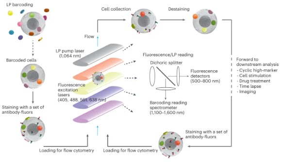 Fig.1 Schematic diagram of multi-pass flow cytometry.1
Our In Vitro Cell-based Analysis Service by Multicolor Flow Cytometry
Creative Biolabs provides a comprehensive, end-to-end solution for multicolor flow cytometry analysis, guiding your project from concept to conclusion. Our services are built upon a foundation of expert consultation and state-of-the-art execution.
In Vitro Immune Cell Population Analysis by Multicolor Flow Cytometry
This service provides a high-resolution snapshot of the cellular composition of your sample. We employ expertly designed panels to delineate major immune lineages and deep-dive into specific subsets, such as T-cell memory and differentiation states (Naïve, Central Memory, Effector Memory, TEMRA), regulatory T-cells, B-cell maturation stages, and the full spectrum of myeloid cells, including monocytes, macrophages, and dendritic cell subsets. This analysis is fundamental for understanding the cellular landscape in homeostasis and disease.
In Vitro Functional and Intracellular Cytokine Analysis by Multicolor Flow Cytometry
Moving beyond phenotypic identification, this service interrogates cellular function. We perform in vitro stimulation of your cells with relevant antigens, mitogens, or antibodies, followed by intracellular staining to measure the production of key effector molecules. This allows for the direct assessment of a cell's functional capacity, such as the polyfunctionality of T-cells (simultaneous production of IFN-γ, TNF-α, IL-2), the degranulation of NK cells (CD107a), or the proliferative capacity of lymphocytes (Ki-67).
Our Advantages
Decades of Expertise
Our team possesses over 20 years of collective experience in high-complexity flow cytometry. We have navigated the evolution of the technology and can overcome the most challenging technical hurdles.
Cutting-Edge Instrumentation
We invest in the latest spectral cytometry and cell sorting technology to provide our clients with the deepest and most accurate cellular insights possible.

True End-to-End Partnership
We function as an extension of your research team, providing dedicated project management and scientific consultation from initial experimental design through to the final data analysis report and publication-ready figures.
Unwavering Commitment to Quality
Our laboratory operates under stringent SOPs and a robust quality management system, ensuring that the data we deliver is accurate, reproducible, and reliable.
FAQs
-
What is the primary difference between conventional and spectral flow cytometry?
Conventional flow cytometry uses a series of optical filters to assign a specific detector to each fluorochrome. Spectral flow cytometry captures the full emission spectrum of light from each cell and uses a complex algorithm to computationally separate the signal from each dye. This allows for more flexibility, higher parameter counts, and better management of cellular autofluorescence.
-
How many markers can I realistically analyze at once?
With our spectral cytometry platforms, analyzing 30-40 markers is routine. For exceptionally complex projects, panels exceeding 50 markers can be designed and executed.
-
What are your sample input requirements?
We can accommodate a wide range of sample types and cell numbers. Requirements vary from assay to assay. Please contact us to discuss the specifics of your project.
-
How do you handle data analysis for high-parameter experiments?
We strongly recommend and provide expertise in computational, algorithm-based analysis for any panel exceeding 15 colors. We provide raw data, gated populations, and advanced visualizations like UMAP plots.
Contact Us
Advance your research with unparalleled cellular insight. To discuss your project with a Creative Biolabs flow cytometry specialist or to request a quote, please contact our project management team.
Reference
- Kwok, Sheldon J J et al. "High-dimensional multi-pass flow cytometry via spectrally encoded cellular barcoding." Nature Biomedical Engineering vol. 8,3 (2024): 310-324. doi:10.1038/s41551-023-01144-9. Distributed under an Open Access License CC BY 4.0, without modification.
For Research Use Only.