Creative Biolabs' spontaneous tumor animal models play an irreplaceable role in oncology research due to their high biological relevance and ability to faithfully simulate the entire natural progression of human tumor development, from initiation to advancement. Our comprehensive platform includes both Conventional Spontaneous Mouse Models and Gene-Edited Spontaneous Mouse Models, significantly broadening the scope and depth of mechanistic and therapeutic investigations. These highly representative spontaneous models are essential for preclinical validation of novel prevention strategies aimed at interrupting the early stages of carcinogenesis in high-risk populations.
Available Spontaneous Tumor Animal Models
Creative Biolabs leverages the superior biological relevance of Spontaneous Tumor Animal Models to provide highly predictive data, moving beyond the limitations of simple transplant models. Conventional Spontaneous Mouse Models develop tumors naturally in their native tissue, providing the most authentic tumor microenvironment (TME) and progression timeline. Ideal for studying cancer initiation, metastatic progression, and assessing long-term drug effects within an immunocompetent host.
Fig1. Spontaneous mutations.1,4
Tumor Type
Incidence Rate (%)
Spontaneous Age (Months)
Modeling Method/Mechanism
Relevant Evaluation
Animal Species
Mammary Carcinoma
80-100
8-12
MMTV Expression/High Susceptibility
Breast Cancer, Hormonal Therapy
C3H
Spontaneous Leukemia
>90
6-12
High MuLV Load/Genetic Predisposition
Hematological Malignancy, Anti-Leukemia Drugs
AKR
Lymphoma
80-90
18-24
Aging/Genetic Susceptibility
Immunotherapy, Aging Research
C57BL/6J
Ovarian Carcinoma
70-80
12-18
Endocrine Imbalance/Failure
Ovarian Cancer, Hormonal Signaling
BALB/c
Pulmonary Adenoma
80-90
12-18
Spontaneous Kras Activation/Genetic
Lung Cancer Prevention, Mutagenesis
A/J
Hepatocellular Carcinoma
70-80
18-24
Hepatitis Virus Susceptibility/Genetic
Liver Cancer, Chronic Inflammation
C3H/He
Adrenocortical Tumors
50-70
12-18
Prone to developing adrenocortical adenomas/carcinomas, associated with endocrine dysfunction.
Endocrine Tumors, Hormone Therapy
NIH/O
Pituitary Tumor/Adenoma
30-94
18-30
High spontaneous rate in aged animals; linked to hormonal/neuroendocrine factors.
Pituitary Tumor Research, Endocrine Drug Screening, Geriatric Toxicology
Wistar Rat, F344/OM Strains
Glandular Stomach Carcinoma
40-60
>18
High incidence of glandular stomach carcinoma upon aging.
Gastric Cancer Pathogenesis, H. pylori Studies
NZO/HlLt
Soft Tissue Sarcoma
30-50
12-24
Spontaneously develops sarcomas and tumors of the reticuloendothelial system.
Sarcoma Research, Cell Signaling Pathways
SJL/J
Endometrial Cancer (EC)
90
>18-24
High incidence in aged females, potentially linked to hormonal dysregulation.
Endometrial/Uterine Cancer Studies, Hormonal Therapies
BDII/Han Rat
Testicular Tumor
86
>24
High incidence in aged males, often Leydig cell tumors.
Testicular Cancer Research, Endocrine Disruptors
F344 Male Rat
Gene-Edited Spontaneous Mouse Models feature precise genetic manipulation (knock-in/knock-out) to drive tumor formation with specific, clinically relevant oncogenic mutations. Offer unparalleled control and translational relevance, allowing to test targeted therapies (like XDCs) against specific, human-relevant drivers (e.g., KRAS mutation, HER2 amplification) in a natural setting. De-risk clinical pipeline by providing robust in vivo proof-of-concept for targeted therapies and combination strategies, ensuring efficacy is tied directly to the intended genetic pathway. Creative Biolabs offers Spontaneous model mice with various targets:
Fig.2 A summary of the similarities detected between mouse mammary tumors and human tumors across different cancer types.2,4
Evaluation Platform
Our comprehensive evaluation platform integrates cutting-edge instrumentation and specialized assays to provide a multi-dimensional view of drug efficacy, pharmacodynamics, and mechanism of action:
Real-Time, Non-Invasive Imaging:
IVIS & High-Resolution Ultrasound: Enables real-time, dynamic, and precise monitoring of tumor burden, metastasis, and angiogenesis, allowing longitudinal tracking of PD effects without culling animals.
Deep Mechanistic and Immune Analysis:
Flow Cytometry (FACS): Provides in-depth profiling of immune cell populations (TILs) within the tumor microenvironment, assessing drug impact on the immune system for both solid and hematological tumors.
Digital Pathology: Uses quantitative IHC/IF to perform objective, high-throughput quantification of biomarkers like Ki-67 and apoptosis markers within tumor tissue.
Molecular and Pharmacodynamic Validation:
qPCR/ddPCR & Western Blot: Validates target engagement and signaling pathway inhibition (p-AKT) at the molecular level, and monitors resistance mutations in plasma (ctDNA).
Survival Analysis: Combined with accurate gross pathology, this determines Overall Survival (OS) and tumor incidence rates, serving as the definitive readout of drug efficacy.
Applications
Mechanistic Insight and Genetic Susceptibility: They serve as ideal tools for revealing carcinogenesis mechanisms and exploring how genetic susceptibility drives malignant transformation.
TME and Immune Interaction: These models genuinely reflect the complexity of the Tumor Microenvironment (TME) and are vital for in-depth studies on the dynamic interaction between the tumor and the immune system.
Long-Term Strategies and Personalized Medicine: Given their longer progression cycle, spontaneous tumors are the foundation for evaluating long-term treatment strategies and chronic drug toxicity. Simultaneously, by simulating distinct human tumor subtypes, they strongly support the development and validation of precision medicine and personalized treatment regimens.
Our Advantages
Extensive experience and professional expertise: We have extensive expertise in tumor research and a profound comprehension of diverse spontaneous tumor models. This foundation enables the delivery of expert research design and specialized scientific guidance.
Diverse model library: We maintain a diverse repository of animal model resources, encompassing a wide range of tumor types and subtypes, ensuring the accommodation of varied research requirements.
Advanced facilities and technical platforms: We are equipped with modern experimental facilities and high-end technical platforms capable of supporting intricate experimental designs and facilitating comprehensive, multi-parameter data analysis.
High-quality data generation and analysis capabilities: We deliver high-standard data collection and analytical services, ensuring the reliability and reproducibility of all resulting research data.
Interdisciplinary collaboration: We are uniquely positioned to foster multidisciplinary collaboration across fields such as medicine, pharmacy, and biology.
Work with Us
1
Inquiry Stage:
Summarize the project requirements and fill in the information collection form.
Sign a CDA from both parties to further communicate information, such as targets.
Select an animal model, discuss experimental design, and determine assay parameters.
Project costing and project schedule forecasting.
2
Project Start:
We provide a detailed project plan, including the required sample quantities, methods, and protocols.
Both parties confirm the project details and start the project.
Confirm the timeline of the project.
3
Project Progress:
We provide periodic results and information on the animal's condition.
We will work together to make project adjustments as necessary.
4
Project Completion:
We provide a comprehensive project report promptly.
We arrange transportation for the produced samples.
We provide a discussion of the project results and help to arrange the next steps.
5
After-Sales Support:
Data storage and archiving.
Get in touch!
FAQs
Q: What are the advantages of spontaneous tumor models?
A: These models offer superior translational fidelity by accurately recapitulating the inherent heterogeneity and natural progression characteristic of human malignancy, alongside the critical interaction with the endogenous host immune system. This fidelity provides a more realistic foundation for assessing therapeutic efficacy and clarifying underlying mechanisms.
Q: What types of spontaneous tumor models are provided?
A: A diverse repository of spontaneous tumor models is provided, encompassing, but not restricted to, common neoplastic entities such as mammary carcinoma, pulmonary neoplasia, colorectal adenocarcinoma, and dermal malignancy.
Q: What types of research can spontaneous tumor animal models be used for?
A: These platforms are instrumental for evaluating the efficacy and tolerability of novel pharmacological agents, conducting detailed investigations into the tumor immune microenvironment, dissecting fundamental tumor biological mechanisms, and facilitating the prediction of human clinical trial outcomes.
Q: What is the typical duration of the experiment?
A: The study duration is contingent upon the specific tumor model and the predefined research objectives, typically ranging from several weeks to multiple months.
Q: How is the reliability of research data ensured?
A: We mandate strict adherence to comprehensive Standardized Operating Procedures (SOPs) and implement rigorous quality control (QC) protocols. This approach ensures the accuracy and consistency of data across both the execution of experiments and subsequent analysis.
Published Data
Fig.3 Tumor growth and lung metastasis of different cells in an in vivo metastasis model.3,4
Using a spontaneous lung metastasis mouse model combined with gene editing technology, it was confirmed that the nicotinic acetylcholine receptor subtype Alpha-9 (α9-nAChR) mediates in vivo tumor growth and lung metastasis of triple-negative breast cancer.
References
Onaciu, Anca et al. "Spontaneous and Induced Animal Models for Cancer Research." Diagnostics (Basel, Switzerland) vol. 10,9 660. https://doi.org/10.3390/diagnostics10090660
Hollern, Daniel P et al. "Histological subtypes of mouse mammary tumors reveal conserved relationships to human cancers." PLoS Genetics vol. 14,1 e1007135. https://doi.org/10.1371/journal.pgen.1007135
Huang, Li-Chi et al. "Nicotinic Acetylcholine Receptor Subtype Alpha-9 Mediates Triple-Negative Breast Cancers Based on a Spontaneous Pulmonary Metastasis Mouse Model." Frontiers in cellular neuroscience vol. 11 336. https://doi.org/10.3389/fncel.2017.00336
Distributed under Open Access license CC BY 4.0, without modification.
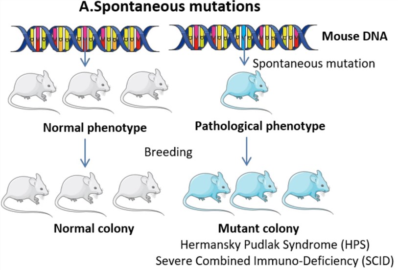 Fig1. Spontaneous mutations.1,4
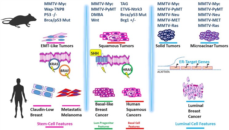 Fig.2 A summary of the similarities detected between mouse mammary tumors and human tumors across different cancer types.2,4
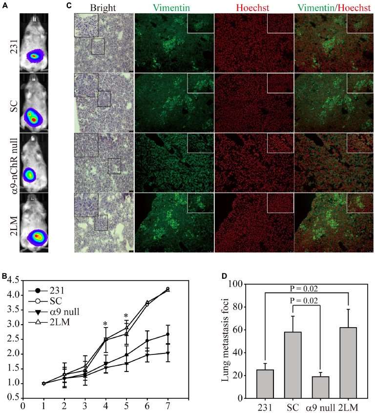 Fig.3 Tumor growth and lung metastasis of different cells in an in vivo metastasis model.3,4