Targeted Molecule-Drug Conjugate (XDC) based Efficacy Modeling & Pharmacodynamics Service
Introduction
Targeted Molecule-Drug Conjugates (TDCs), also known as XDCs, are a broad class of therapeutic drugs that use a targeting molecule, such as a monoclonal antibody, small molecule, or peptide, corresponding to types like Antibody-Drug Conjugates (ADCs), Small Molecule-Drug Conjugates (SMDCs), Bispecific ADCs (BsADC), DNA-natural product conjugate, Antibody–biopolymer conjugate (ABC), Oligonucleotide-Drug Conjugates (ODCs), Degradative-Antibody conjugate (DACs), and Peptide-Drug Conjugates (PDCs), to deliver potent drugs to specific cells. Initially reliant on a single cytotoxic payload, TDCs now integrate immune modulation, bispecific synergy, and smart-release mechanisms to balance efficacy with toxicity. With breakthroughs in technologies like bispecific ADCs, TDCs are set to expand into cutting-edge fields such as gene editing and protein degradation, ushering in a new era of multi-modal, precision medicine for cancer treatment. By precisely understanding the characteristics, pros and cons, and mechanisms of action of XDCs, Creative Biolabs selects appropriate animal models to evaluate their preclinical pharmacology, toxicology, and pharmacokinetics, thereby accelerating their development process.
Fig.1 The cytotoxic drugs (payloads) used for XDCs typically have the characteristics listed above.
Available XDCs Types Efficacy Services
XDCs Types
Targeted Molecule
Modeling & Applications
ADCs
Monoclonal Antibody (mAb)
ADCs are primarily screened in CDX/PDX models, and their immune mechanisms (ADCC/ADCP) and IO synergy are evaluated in Syngeneic/HSC Humanized animal models.
SMDCs
Small Molecule Ligand (e.g., Folate, PSMA inhibitor)
SMDCs utilize In Vivo PET/MRI imaging to track rapid PK/PD characteristics and employ NHP models to assess organ toxicity stemming from off-target small molecule ligand binding.
BsADC
Bispecific Antibody (BsAb) (Targets two distinct antigens)
BsADCs utilize GEMMs (engineered to express dual targets) to evaluate efficacy against target heterogeneity and rely on HSC Humanized models to enhance T/NK cell immune activation.
PDCs
Peptide (short chain of amino acids)
PDCs require critical PK/PD analysis (LC-MS/MS) in animal models to optimize dosing schedules due to rapid clearance, and are used to verify superior in vivo tumor penetration compared to ADCs.
ABC
Aptamer (Single-stranded nucleic acid or peptide molecule)
ABCs primarily use NHP models to assess the systemic safety and PK characteristics of the nucleic acid Aptamer, while animal models monitor its overall stability.
ODCs
Deoxyribonucleic Acid (DNA) or Oligonucleotide (Less common usage)
ODCs utilize Gene-Edited animal models to evaluate the therapeutic efficacy of the nucleic acid payload, with fluorescently labeled XDCs tracking in vivo delivery and accumulation.
DACs
Monoclonal Antibody (mAb) linked to a Protein Degrading Moiety
DACs are evaluated primarily in CDX/PDX models to confirm in vivo target protein degradation, with GEMMs offering the potential to provide mechanistic proof of sustained knockdown in spontaneous tumors.
Fig.2 Key components and mechanisms of action of ADCs.2,3
Animal models are a crucial part of the preclinical evaluation of TDCs, bridging the gap between lab based research and human clinical trials. Creative Biolabs is professionalized in antibody-associated drug discovery and has versatile in vivo platforms for TDCs development, the most widely used animal models for TDCs evaluation are rodent models, primarily mice. These can be categorized into a few types:
Xenograft mouse models: Established tumor bank of cell line derived xenografts (CDXs) for accurate selection of targets. Patient derived xenografts (PDXs) for modeling metastases and tumor heterogeneity. Models customizable with human immune system reconstitution and human immune checkpoint knock-in mice.
Syngeneic mouse models: A common challenge is that XDCs are usually designed to target human antigens (targets), which are not expressed by the murine syngeneic tumor cells. To overcome this, we employ two key strategies:
Engineered Syngeneic Models: Humanized Mouse Tumor Cell Lines are established by genetically modifying murine tumor lines (e.g., CT26, 4T1) to stably express the human target antigen (e.g., human BCMA, CCR4, CD19, CD38, CD47, CEA, CEACAM1, CLDN18.2, CXCR2, CXCR4, EPCAM, HER2, PD-L1, PD-L2, ROR1, TNFR2, Trop-2). These modified cells are then implanted into the original, immune-competent mouse strain. This allows the testing of the clinical candidate XDC (the human-targeting drug) in an immunocompetent environment. Furthermore, we use Fluorescence labeling cell lines to visualize, quantify, and track the XDC at the cellular and tissue level, addressing critical questions about its efficacy and pharmacodynamics (PD).
Surrogate ADCs: Alternatively, a surrogate XDC targeting the murine homolog of the antigen can be used in the unmodified syngeneic model.
Genetically Engineered Mouse Models (GEMMs): By modeling spontaneous human cancers with key drivers (e.g., HER2 overexpression, KRAS mutation, or TP53 loss), Humanized Immune System Mice Models (e.g., Humanized Immune Checkpoint Models, Humanized Tumor Target Models), GEMMs provide a complete, immune-competent tumor microenvironment. This unique capability is vital for predicting clinical success, accurately validating the XDC's mechanism, and proving the synergy of combination therapies. Move beyond traditional models and gain confidence in your drug candidates.
Humanized Immune Models (PBMC and HSC): These models are essential for XDC success. We provide a functional human immune system in vivo to validate your drug's precise mechanism, confirm critical Fc-effector functions (ADCC/ADCP), and prove synergy with immunotherapy. Use our most predictive platforms to de-risk your XDC pipeline and achieve faster clinical translation.
Tumor Organoid Models: Tumor Organoid Models are your most predictive 3D platform for XDC success, uniquely enabling you to validate target penetration and confirm the mechanism of action, from Target-Binding to full 3D cytotoxicity, all while rapidly predicting patient response across diverse tumor profiles. For example, patient derived organoids (PDxOs) are rapidly generated from patient samples for HTS. They pinpoint the best XDCs for specific genotypes (e.g., HER2 overexpression, KRAS mutation). As PDxO response predicts clinical outcomes, they are indispensable for prioritizing XDC candidates.
Drug-resistant models: These models are your safeguard against clinical failure, allowing you to proactively identify and overcome tumor escape mechanisms, such as target downregulation (e.g., HER2 loss) or payload efflux, to design next-generation XDCs with superior, durable efficacy.
Preclinical evaluation: Our advanced detection methods provide unparalleled insight into XDC performance, utilizing Flow Cytometry for detailed cellular characterization, and Confocal Microscopy with Lyso-Tracker staining to directly confirm lysosomal endocytosis and payload delivery. We monitor in vivo efficacy using both IVIS imaging and PET-CT dynamic monitoring for early metabolic response assessment, while IHC and RNA in situ hybridization characterize the critical Tumor Microenvironment (TME) for combination strategy guidance. Furthermore, we precisely define drug stability and quality through LC-MS/MS and immunoassay for PK analysis, and Immuno-enrichment coupled with mass spectrometry for dynamic tracking of the essential Drug-to-Antibody Ratio (DAR). We deliver comprehensive predictive analysis, integrating efficacy, toxicity, PK, and biomarker data to forecast individualized patient response. By establishing the quantitative relationship between drug concentration, time, and resulting effect (C-T-E), we accurately predict the duration and strength of therapeutic effect for various doses. This ultimately accelerates the research and development timeline and boosts the success rate of clinical translation.
Fig.3 A four-dimensional evaluation system.
Applications
Preclinical Efficacy Assessment & Screening: We establish a quantitative correlation between in vitro cell killing activity and in vivo animal model efficacy to quickly eliminate low-efficiency candidates during the drug discovery stage.
Tumor Model Efficacy Prediction: We evaluate the relationship between target expression and drug efficacy to screen for highly responsive tumor types. This process helps predict individualized treatment response and guides biomarker development.
PK Feature Analysis: We establish a "concentration-time-effect" quantitative relationship to predict the duration and intensity of drug efficacy across different dosage levels.
Safety and Toxicity Prediction: We construct models of the normal tissue target expression-drug concentration-toxicity relationship to predict the potential risk of damage to organs such as the liver and kidney, guiding the selection of linkers and payloads. Furthermore, we incorporate Anti-Drug Antibody (ADA) kinetic models to predict the risk of efficacy decline during long-term dosing.
Our Advantages
One-Stop Service: The platform offers a seamless, one-stop solution for targeted drug development, combining an extensive model library with advanced analytical capabilities and proven clinical expertise.
Integrated Model Development: Possess end-to-end model construction capabilities, ranging from in vitro to in vivo and comprehensive Pharmacokinetic (PK) to Pharmacodynamic (PD) assessment.
Integrated Platform: Access our comprehensive library of 300+ validated CDX/PDX models covering highly relevant targets, including HER2, TROP2, and Claudin 18.2. Proven specialized experience in evaluating various cancer types, adept at managing both solid tumors and hematological malignancies.
Advanced Detection Methods: Utilizing cutting-edge techniques like imaging (e.g., PET, SPECT, MRI), molecular analysis (e.g., qPCR, NGS, Mass Spectrometry), and cellular/histological assays (e.g., IHC, Flow Cytometry) for the comprehensive and precise in vivo monitoring of tumor characteristics, drug mechanism of action, and cell populations.
Extensive Experience: We have a proven track record, having supported the clinical approval of multiple XDC drug candidates, ensuring translationally relevant results.
Complete Bioanalytical Capability: Equipped to perform full XDC component bioanalysis, covering the total drug, free payload, and metabolites.
Customized Modeling: Ability to provide exclusive model solutions based on the specific properties of your drug, such as linker type and payload characteristics.
Work with Us
1
Inquiry Stage:
Summarize the project requirements and fill in the information collection form.
Sign a CDA from both parties to further communicate information, such as targets.
Select an animal model, discuss experimental design, and determine assay parameters.
Project costing and project schedule forecasting.
2
Project Start:
We provide a detailed project plan, including the required sample quantities, methods, and protocols.
Both parties confirm the project details and start the project.
Confirm the timeline of the project.
3
Project Progress:
We provide periodic results and information on the animal's condition.
We will work together to make project adjustments as necessary.
4
Project Completion:
We provide a comprehensive project report promptly.
We arrange transportation for the produced samples.
We provide a discussion of the project results and help to arrange the next steps.
5
After-Sales Support:
Data storage and archiving.
Get in touch!
FAQs
Q. How are TDCs different from traditional chemotherapy?
A: Traditional chemotherapy indiscriminately kills fast-dividing cells, resulting in damage to healthy tissue, whereas Targeted Molecule-Drug Conjugates (TDCs) function as "smart missiles," selectively binding to specific targets on cancer cells and releasing the drug only upon internalization, thereby minimizing damage to healthy tissue and significantly reducing side effects.
Q. What are the key considerations when choosing an animal model?
A: Selecting the right model is crucial for obtaining meaningful data, requiring researchers to carefully consider several factors: Target Expression must be high and relevant to the TDC's design; Tumor Type & Location should mimic the human disease as closely as possible; the Immune System must be chosen based on the research question, necessitating an immunocompetent model (like syngeneic) if the TDC's effect on immunity is key; and finally, Toxicity must be assessed using a susceptible species, potentially requiring a non-human primate model if the target is expressed in primates but not rodents.
Q. Besides tumor size, what other indicators are observed to assess TDC efficacy in animal models?
A: To comprehensively evaluate a TDC's effectiveness, we monitor detailed indicators beyond simple tumor size, including tracking Biomarker Changes and Pharmacodynamic (PD) Markers to confirm the drug's expected biological effects and downstream signaling changes within tumor cells. We also assess In Vivo Distribution using imaging techniques like PET or MRI to confirm effective accumulation at the tumor site, and evaluate Tumor Microenvironment Changes (involving blood vessels, immune cells, and stromal components) as these are critical factors linked to successful treatment outcomes.
Q. How are dosing and administration schedules determined in TDC animal studies?
A: Determining the correct dose and schedule is crucial and is typically based on Dose-Escalation Studies to establish the Maximum Tolerated Dose (MTD) and identify toxicities. This is further refined by PK/PD-Guided Dosing, which utilizes pharmacokinetic (drug concentration) and pharmacodynamic (biological effects) data to ensure the TDC remains within an effective, sustained concentration window. Finally, Multiple Dosing Regimens are employed to mimic clinical schedules and assess cumulative toxicity and long-term efficacy.
Q. How does data from animal models support a TDC's first-in-human trial?
A: Data derived from animal models is essential for supporting clinical development by informing the Determining the Starting Dose, calculating a safe and effective initial human dose based on pharmacokinetics, efficacy, and toxicity. This information is also crucial for Predicting Potential Toxicities observed in animals, allowing researchers to establish necessary monitoring and management plans. Furthermore, animal studies aid in establishing the Dosing Schedule for clinical protocols and provide vital Evidence of the mechanism of Action through in vivo biomarker data, enhancing the overall understanding of the TDC's potential effects in humans.
Published Data
Fig.4 A Structures of anti-HER2 AF488/Cy5 dual-dye conjugate (DOL of 2 + 2) and single-dye variants (DOL of 2). Fluorescence images of the whole tumors (b) and semi-quantification (c) detected using a 700 nm channel. d–f Fluorescence microscopic imaging of the tumor tissues.2,3
Evaluation of payload delivery efficiency by the dual conjugate format: In the case of a single-payload combination therapy, there is competition for the HER2 target, which reduces delivery efficiency. However, dual-payload ADCs do not have this issue.
References
Shi, Ruotong et al. "Another power of antibody-drug conjugates: immunomodulatory effect and clinical applications." Frontiers in immunology vol. 16 1632705. https://doi.org/10.3389/fimmu.2025.1632705
Yamazaki, Chisato M et al. "Antibody-drug conjugates with dual payloads for combating breast tumor heterogeneity and drug resistance." Nature communications vol. 12,1 3528. https://doi.org/10.1038/s41467-021-23793-7
Distributed under Open Access license CC BY 4.0, without modification.
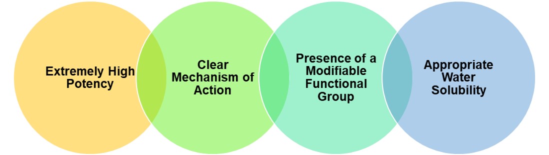 Fig.1 The cytotoxic drugs (payloads) used for XDCs typically have the characteristics listed above.
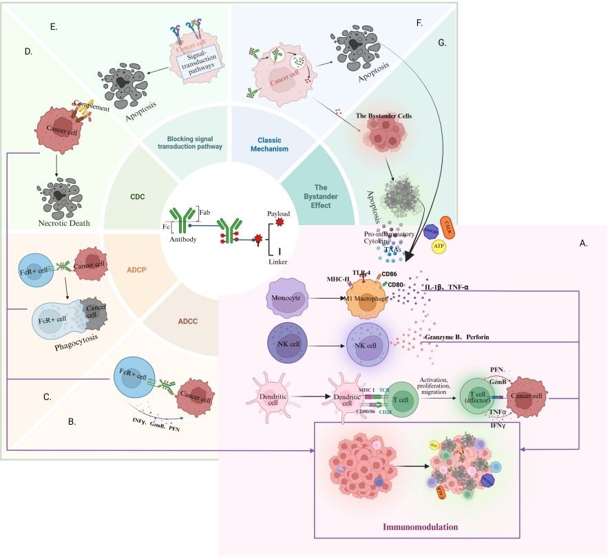 Fig.2 Key components and mechanisms of action of ADCs.2,3
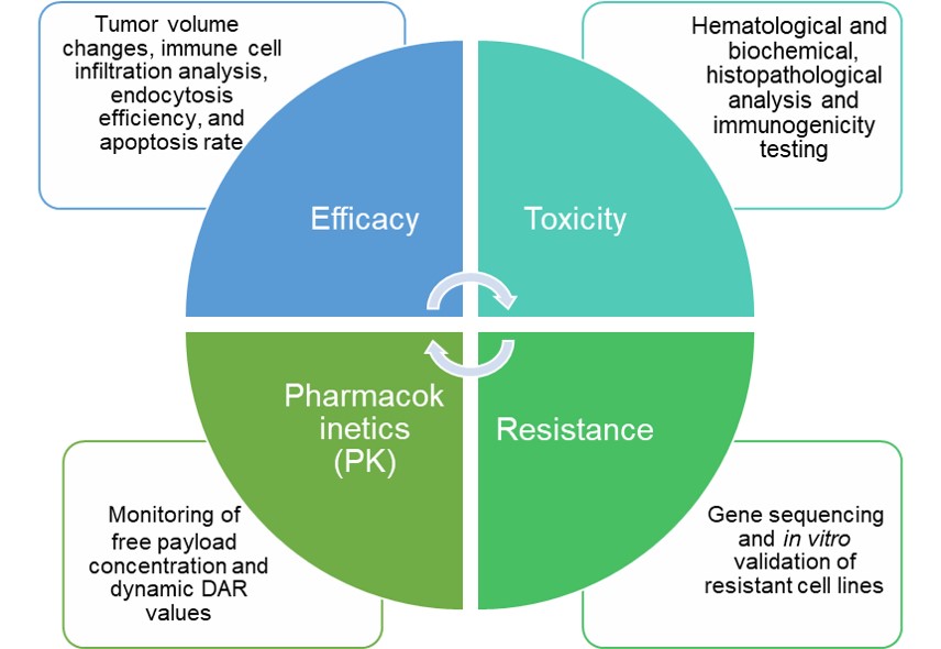 Fig.3 A four-dimensional evaluation system.
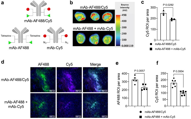 Fig.4 A Structures of anti-HER2 AF488/Cy5 dual-dye conjugate (DOL of 2 + 2) and single-dye variants (DOL of 2). Fluorescence images of the whole tumors (b) and semi-quantification (c) detected using a 700 nm channel. d–f Fluorescence microscopic imaging of the tumor tissues.2,3