
Anterior Cruciate Ligament Transection (ACLT) induced Osteoarthritis Modeling & Pharmacodynamics Service
Are you currently facing high variability in joint degradation models, inconsistent histopathological scoring, or challenges in translating in vitro results to clinical outcomes? Our ACLT-Induced Osteoarthritis Modeling & Pharmacodynamics Service helps you validate therapeutic efficacy and obtain high-quality longitudinal data through surgical precision and advanced molecular phenotyping. Creative Biolabs accelerates your R&D timeline using standardized surgical protocols and multi-dimensional analysis platforms tailored for disease-modifying osteoarthritis drugs.
Overview of ACLT-Induced Osteoarthritis Modeling
The Anterior Cruciate Ligament (ACL) is the primary stabilizer of the knee joint. Its rupture, either through acute trauma or surgical transection, leads to immediate mechanical instability, altered joint loading, and a cascade of biochemical changes. ACLT-induced Osteoarthritis (OA) is the premier model for studying Post-Traumatic Osteoarthritis (PTOA). Unlike chemical or aging models, ACLT provides a clear timeline of disease progression, starting with an acute inflammatory phase followed by chronic cartilage erosion, subchondral bone remodeling, and osteophyte formation. This model is essential for researchers targeting early-stage interventions and the prevention of vascular invasion into the articular matrix.
ACLT-Induced Osteoarthritis Models
Creative Biolabs provides highly standardized ACLT models in both the rat and Rabbit species. Our surgical team utilizes microsurgical techniques to ensure minimal collateral tissue damage while achieving consistent joint instability. These models are validated to demonstrate hallmark OA features, including synovial hyperplasia, chondrocyte apoptosis, and extracellular matrix (ECM) degradation, within a predictable 4-to-12-week window. Our protocols are optimized to provide low inter-animal variability, ensuring your pharmacodynamic studies achieve high statistical power with smaller cohort sizes.
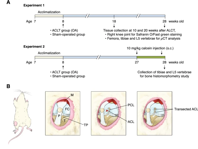 Fig.1 Illustrations depict the procedural sequence of the knee osteoarthritis (OA) model induced by ACLT.1,3
Evaluation Platform of Our Service
Creative Biolabs offers a comprehensive suite of analytical tools to quantify the efficacy of your therapeutic candidates. Our platform integrates molecular precision with functional outcomes:
-
Biochemical & Molecular Analysis:
- Cytokine Profiling: Multiplex assays in synovial fluid.
- Catabolic Enzymes: qPCR and Western Blot.
- Biomarker Tracking: ELISA for CTX-II (Collagen Type II degradation) and COMP.
-
Histopathological Examination:
- Tissue Staining: Safranin-O/Fast Green, Hematoxylin & Eosin (H&E), and Toluidine Blue.
- Scoring Systems: Standardized OARSI and Mankin scoring by blinded pathologists.
- Immunohistochemistry (IHC): Localization of VEGF-A, Type II Collagen, and Caspase-3.
-
Behavioral & Functional Testing:
- Gait Analysis: Automated footprint analysis to measure stride length and pressure.
- Weight Bearing: Incapacitance testing to measure limb-specific pain distribution.
- Motor Coordination: Rota-rod and open-field activity monitoring.
-
Advanced Imaging & Instrumentation:
- Micro-CT: High-resolution subchondral bone volume and osteophyte quantification.
- MRI: Non-invasive T2 mapping for cartilage hydration and structural integrity.
- Bone Mineral Density (BMD): Measurement of subchondral plate thickening.
Key Applications
Our ACLT models are versatile tools for the pharmaceutical and biotech industries, simulating various stages of joint disease:
- Indications: Post-traumatic osteoarthritis, chronic joint inflammation, subchondral bone sclerosis, and meniscus-related secondary degradation.
- Therapeutic Agents: Small Molecules: NSAIDs, kinase inhibitors, and mitochondrial stabilizers.
- Biologics: Monoclonal antibodies (anti-VEGF, anti-NGF), growth factors (TGF-β), and recombinant proteins.
- Gene Therapies: AAV-mediated delivery of chondroprotective genes.
- Cell-Based Therapies: MSC-based regeneration and exosome-derived treatments.
Why Choose Us?
Partnering with Creative Biolabs means gaining access to a specialized ecosystem designed for orthopedic success:
We offer fully validated strains of Rat and Rabbit, allowing for scalability from early-stage screening to larger-tissue surgical evaluations.
We manage the entire pipeline, from surgical induction and dosing (intra-articular, systemic, or topical) to final histopathological reporting.
Our team consists of PhD-level biologists and veterinary surgeons with decades of experience in musculoskeletal research, supported by a rigorous ISO-compliant quality management system.
Work with Us
- Summarize the project requirements and fill in the information collection form.
- Sign a CDA from both parties to further communicate information, such as targets.
- Select an animal model, discuss experimental design, and determine assay parameters.
- Project costing and project schedule forecasting.
- We provide a detailed project plan, including the required sample quantities, methods, and protocols.
- Both parties confirm the project details and start the project.
- Confirm the timeline of the project.
- We provide periodic results and information on the animal's condition.
- We will work together to make project adjustments as necessary.
- We provide a comprehensive project report promptly.
- We arrange transportation for the produced samples.
- We provide a discussion of the project results and help to arrange the next steps.
- Data storage and archiving.
Frequently Asked Questions
-
Q: How soon after ACLT surgery can we begin dosing our therapeutic candidate?
A: Dosing typically begins between Day 1 and Week 2 post-surgery, depending on whether you are targeting the acute inflammatory phase or chronic degradation. We can customize the initiation based on your drug's mechanism of action.
-
Q: Do you offer intra-articular (IA) injection services for Rat and Rabbit models?
A: Yes, Creative Biolabs specializes in precise IA injections. Our surgeons use ultrasound guidance or anatomical landmarks to ensure the therapeutic agent is delivered directly into the synovial space without damaging the joint capsule.
-
Q: Can your platform distinguish between symptomatic relief and disease modification?
A: Absolutely. By combining behavioral pain testing (symptomatic) with micro-CT and OARSI scoring (disease-modifying), we provide a clear picture of how your agent affects both patient-centric outcomes and structural integrity.
-
Q: What is the typical study duration for an ACLT pharmacodynamics study?
A: Most studies run between 4 and 12 weeks. Short-term studies (4-6 weeks) are ideal for anti-inflammatory screening, while long-term studies (8-12 weeks) are better for observing cartilage protective effects.
Published Data
Objective: To compare the efficacy of regenerative biologics versus traditional corticosteroids in a standardized ACLT model.
Model Used: Rat ACLT (Post-Traumatic Osteoarthritis Model).
Results: The ACLT model effectively demonstrated superior sensitivity in distinguishing long-term outcomes between treatments. While initial pain relief was observed across cohorts, the model's progressive pathology revealed significant joint space narrowing and elevated Mankin scores in the steroid group by Week 12. Conversely, it successfully captured the structural preservation of the regenerative group, characterized by maintained Type II Collagen and a 35% reduction in ADAMTS5. This validation underscores the model's reliability for longitudinal screening, proving PRP-like biologics offer superior structural durability over corticosteroids.
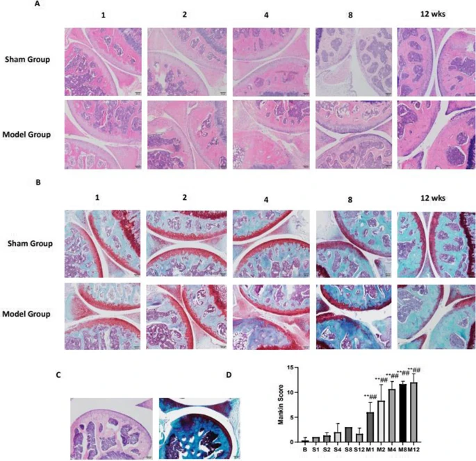 Fig.2 Microscopic examination of cartilage samples from various groups illustrating progression in the ACLT model.2,3
Creative Biolabs is dedicated to providing the high-fidelity data you need to move your joint health candidates from the bench to the clinic. Our integrated ACLT modeling and evaluation platform ensures that every study is a step toward a breakthrough. Contact Our Team for More Information and to Discuss Your Project.
References
- Namhong, Supitra, et al. "Knee osteoarthritis in young growing rats is associated with widespread osteopenia and impaired bone mineralization." Scientific Reports 10.1 (2020): 15079. DOI: https://doi.org/10.1038/s41598-020-71941-8.
- Qian, Jia-jia, et al. "Expression of VEGF-A signaling pathway in cartilage of ACLT-induced osteoarthritis mouse model." Journal of orthopaedic surgery and research 16.1 (2021): 379. DOI: https://doi.org/10.1186/s13018-021-02528-w.
- Distributed under Open Access license CC BY 4.0, without modification.
For Research Use Only.