
Femoral Fracture Modeling & Pharmacodynamics Service
Are you currently facing high data variance in bone healing studies, difficulty in maintaining joint integrity during pinning, or challenges in quantifying subtle pharmacological effects on callus formation? Our Femoral Fracture Modeling Service helps you accelerate orthopedic drug discovery and validate mineral-density interventions through surgical mastery and high-resolution analytical platforms, ensuring your therapeutic leads are tested against the highest biological standards.
Overview of Femoral Fracture Modeling
Femoral fractures represent a critical subset of musculoskeletal trauma with high socio-economic impact. The physiological system of bone healing is a complex, multi-phase cascade involving initial hematoma formation, inflammatory signaling, fibrocartilaginous (soft) callus development, and long-term mineralized (hard) callus remodeling. Diseases such as osteoporosis, diabetes, and age-related osteopenia significantly impair this regenerative capacity, often leading to non-union or delayed union. Creative Biolabs provides sophisticated Rat models that simulate these pathological environments, allowing for the rigorous testing of agents designed to stimulate osteoprogenitor recruitment and accelerate structural bridging.
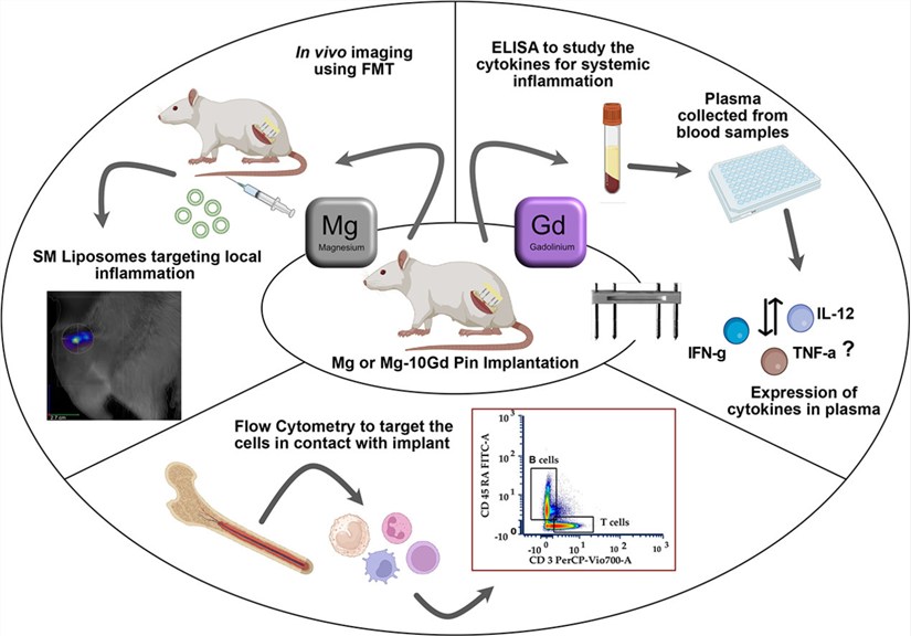 Fig.1 Inflammatory reaction to magnesium-based metallic biomaterial in a rat femur fracture model.1
Femoral Fracture Models
Creative Biolabs offers a range of validated Rat models, including the industry-standard refined antegrade (trochanteric) pinning protocols. These models are specifically designed to preserve the periosteum and surrounding soft tissue, ensuring a natural endochondral ossification response. We offer specialized cohorts, such as Ovariectomized (OVX) Rats to simulate postmenopausal osteoporosis and metabolic-challenge models for diabetic fracture repair.
Evaluation Platform of Our Service
Our platform provides the analytical depth required to move beyond simple visual union, offering a technical parameter list that ensures regulatory-grade data:
-
Biochemical & Molecular Analysis:
- Systemic and local Cytokine profiling (IL-6, TNF-α, TGF-β).
- Western Blot and qPCR for osteogenic markers (Runx2, Osteocalcin, ALP).
-
Histopathological Examination:
- H&E and Goldner's Trichrome staining for soft/hard callus differentiation.
- Standardized Histomorphometry and qualitative scoring systems for bony union.
- Immunohistochemistry (IHC) for vascularization and collagen typing.
-
Behavioral & Functional Testing:
- Automated Gait Analysis to track functional recovery and weight-bearing.
- Grip strength and motor coordination assessments.
- Nash Equilibrium deviation analysis for cognitive-behavioral phenotyping.
-
Advanced Imaging & Instrumentation:
- High-resolution Micro-CT for microarchitectural analysis (BV/TV, Trabecular thickness).
- Dual-Energy X-ray Absorptiometry (DEXA) for Bone Mineral Density (BMD).
- Ex vivo Biomechanical Torsional and 3-point bending tests.
Key Applications
Creative Biolabs' models are utilized to simulate a wide range of indications, including:
- Standard traumatic fracture repair and non-union scenarios.
- Osteoporotic fragility fractures and impaired metabolic healing.
- Critical-sized defects and bone graft performance.
- We evaluate a diverse array of therapeutic modalities, including small molecule osteoanabolics, biologics (growth factors), gene therapies targeting localized expression, and cell-based therapies for regenerative medicine.
Why Choose Us?
We provide access to a wide range of validated Rat strains tailored to your study's specific metabolic or genetic requirements.
Creative Biolabs offers a seamless transition from in vitro screening to in vivo PD/PK studies, providing a unified data stream for your development team.
Our PhD-level team brings over 20 years of experience in orthopedic surgery and behavioral science, supported by a rigorous quality management system that ensures data reproducibility and ethics.
Work with Us
- Summarize the project requirements and fill in the information collection form.
- Sign a CDA from both parties to further communicate information, such as targets.
- Select an animal model, discuss experimental design, and determine assay parameters.
- Project costing and project schedule forecasting.
- We provide a detailed project plan, including the required sample quantities, methods, and protocols.
- Both parties confirm the project details and start the project.
- Confirm the timeline of the project.
- We provide periodic results and information on the animal's condition.
- We will work together to make project adjustments as necessary.
- We provide a comprehensive project report promptly.
- We arrange transportation for the produced samples.
- We provide a discussion of the project results and help to arrange the next steps.
- Data storage and archiving.
Frequently Asked Questions
-
Q: How do you control for comminution variability in your fracture models?
A: We utilize high-resolution intraoperative fluoroscopy to detect and categorize slight comminution. Since comminution is statistically correlated with increased early callus formation, we ensure balanced group allocation to prevent data skewing.
-
Q: Can your platform measure functional recovery in addition to structural healing?
A: Absolutely. We integrate automated gait analysis and motor coordination testing to correlate physical bone bridging with the actual return of function in the Rat.
-
Q: Do you offer models for impaired healing, such as diabetic or aged populations?
A: Yes, we provide specialized Rat cohorts, including aged and metabolically challenged groups, to simulate the high-risk patient populations most relevant to current drug development.
-
Q: What is the typical turnaround time for a 35-day healing study?
A: Most studies are completed within 6-8 weeks from animal acclimation to final histopathological report, though we encourage early inquiry to secure surgical scheduling.
Published Data
Objective: To validate the surgical reliability and functional healing progression of a standardized closed femoral fracture model.
Model Used: Sprague-Dawley Rats subjected to mid-shaft femoral fracture via a blunt-guillotine drop tower, stabilized with intramedullary K-wires using a trochanteric entry approach.
Results: This model ensures high surgical fidelity, with 88% successful stabilization confirmed by fluoroscopy. Quantitative longitudinal analysis demonstrates a predictable endochondral ossification pathway. Crucially, the model captures the biological impact of minor comminution, observed in 63% of cases, which significantly accelerates early soft callus expansion (p=0.015). By standardizing mechanical stability and trauma induction, this platform minimizes confounding variables, providing a high-sensitivity environment for precisely decoupling pharmacological therapeutic effects from intrinsic fracture-healing variability.
Creative Biolabs is your premier partner for high-fidelity Femoral Fracture Modeling & Pharmacodynamics. From surgical mastery to advanced biomechanical validation, we provide the evidence your innovation deserves. For detailed protocols, pricing, or a scientific consultation: Contact Our Team for More Information and to Discuss Your Project.
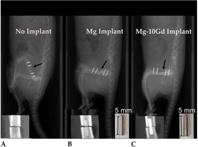 Fig.2 Postsurgical in vivo X-ray imaging across three cohorts.1
Reference
- Riyaz, Sana, et al. "Inflammatory response toward a Mg-based metallic biomaterial implanted in a rat femur fracture model." Acta Biomaterialia 185 (2024): 41-54. Distributed under Open Access license CC BY 4.0, without modification. DOI: https://doi.org/10.1016/j.actbio.2024.06.040.
For Research Use Only.