
Mandibular Defect Modeling & Pharmacodynamics Service
Are you currently facing challenges in bone tissue engineering, such as inconsistent animal models, slow mineralization in large segmental defects, or difficulty in validating scaffold-free constructs? Our Mandibular Defect Modeling & Pharmacodynamics Service helps you obtain high-fidelity preclinical data and optimize therapeutic candidates through validated critical-sized defect (CSD) protocols, advanced Bio 3D-printing integration, and endochondral ossification pathways. We streamline your R&D via rigorous internal fixation techniques and multi-dimensional bone quality analysis.
Overview of Mandibular Defect Modeling & Pharmacodynamics Service
The mandible is a complex physiological system essential for mastication, speech, and facial structural integrity. It is unique among skeletal tissues due to its developmental origin in the neural crest and its constant exposure to multi-axial biomechanical stresses. Diseases and conditions affecting this system, ranging from severe trauma and osteoradionecrosis to extensive tumor resections, often result in segmental defects that exceed the body's natural regenerative capacity. These "critical-sized" gaps necessitate the use of advanced biomaterials, growth factors, or cellular therapies to restore function and aesthetics. Understanding the pharmacodynamics of these interventions requires models that precisely replicate the human clinical environment.
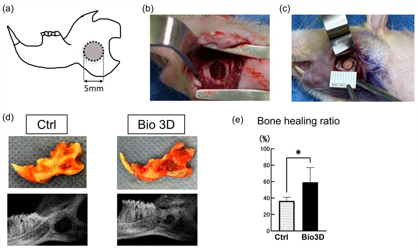 Fig.1 Workflow of a therapeutic method using a mandibular defect model.1
Mandibular Defect Models
Creative Biolabs provides validated Mandibular Defect Models utilizing Rat and Rabbit species. Our primary models include the 5-mm segmental mandibulectomy, established as a definitive critical-sized defect (CSD) that exhibits 0% spontaneous healing without intervention. We also offer specialized alveolar ridge augmentation models and tooth extraction socket models. Each model utilizes rigid internal micro-plating or customized fixation to neutralize masticatory forces, ensuring that observed bone volume increases are a direct result of the test article's therapeutic performance.
Evaluation Platform of Our Service
To demonstrate the depth of our platform, Creative Biolabs offers a comprehensive technical parameter list for evaluating bone quality and regenerative kinetics:
-
Biochemical & Molecular Analysis:
- Osteogenic marker quantification via qPCR.
- Systemic and local cytokine profiling (IL-6, TNF-α, BMP-2) using ELISA.
- Western Blot analysis for signaling pathway activation.
-
Histopathological Examination:
- Standardized staining: H&E, Masson's Trichrome, and Goldner's Trichrome for mineralized vs. non-mineralized tissue.
- Immunohistochemistry (IHC) for vascular markers (CD31, VEGF) and bone-specific proteins.
- Histomorphometric scoring systems for osseointegration and graft degradation.
-
Behavioral & Functional Testing:
- Masticatory efficiency assessments and body weight monitoring post-surgery.
- Grip strength and general motor coordination to ensure post-surgical welfare in Rat and Rabbit models.
-
Advanced Imaging & Instrumentation:
- High-resolution Micro-CT: Measurement of Bone Volume/Total Volume (BV/TV), Trabecular Thickness (Tb.Th), and Trabecular Number (Tb.N).
- Dual-Energy X-ray Absorptiometry (DEXA): Bone Mineral Density (BMD) measurement.
- Biomechanical Testing: Three-point bending and compression tests to evaluate the structural integrity of the regenerate.
Key Applications
Our models effectively simulate a variety of clinical indications, including:
- Segmental Bone Loss: Replicating large gaps resulting from oncological resection or high-impact trauma.
- Osteoporosis-Related Bone Defects: Utilizing specialized strains to study impaired healing environments.
- Alveolar Ridge Atrophy: Testing materials for dental implant site preparation.
These models are suitable for evaluating a broad range of therapeutic agents:
- Small Molecules: Osteoinductive compounds and systemic anti-resorptive drugs.
- Biologics: Recombinant growth factors and targeted antibodies.
- Gene & Cell-Based Therapies: DPSC-derived constructs, ASC-based grafts, and viral vector-mediated gene delivery.
Why Choose Us?
Creative Biolabs focuses exclusively on high-fidelity Rat and Rabbit strains. Each model is pre-validated with historical data to ensure predictable control groups and robust statistical power.
We provide a seamless transition from initial in vitro biocompatibility and cellular differentiation assays to complex in vivo PD/PK studies. Our integrated workflow reduces logistical delays and ensures data continuity across all development phases.
Our team is led by PhD-level scientists with decades of experience in craniofacial surgery and tissue engineering. Supported by a rigorous quality management system, Creative Biolabs guarantees reproducible results that meet the highest standards of international regulatory submissions.
Work with Us
- Summarize the project requirements and fill in the information collection form.
- Sign a CDA from both parties to further communicate information, such as targets.
- Select an animal model, discuss experimental design, and determine assay parameters.
- Project costing and project schedule forecasting.
- We provide a detailed project plan, including the required sample quantities, methods, and protocols.
- Both parties confirm the project details and start the project.
- Confirm the timeline of the project.
- We provide periodic results and information on the animal's condition.
- We will work together to make project adjustments as necessary.
- We provide a comprehensive project report promptly.
- We arrange transportation for the produced samples.
- We provide a discussion of the project results and help to arrange the next steps.
- Data storage and archiving.
Frequently Asked Questions
-
Q: Can you accommodate long-term studies to observe scaffold degradation?
A: Yes, we offer longitudinal studies ranging from 4 weeks to 6 months. Our Micro-CT and histology platforms are specifically designed to track the synchronization of scaffold degradation with new bone formation.
-
Q: What is the primary advantage of using Rabbit models over Rats for certain studies?
A: Rabbit models allow for larger defect volumes and are often preferred for testing the surgical handling properties of bone putties or larger 3D-printed scaffolds. We can recommend the best species based on your specific test article.
-
Q: How do you handle the delivery of fragile cell-based therapies?
A: Our team is experienced in handling scaffold-free 3D constructs and hydrogel-encapsulated cells. We maintain strict cold-chain and specialized surgical protocols to ensure maximum cell viability upon implantation.
-
Q: Is it possible to combine your modeling with systemic PK analysis?
A: Absolutely. We can perform concurrent blood sampling and tissue distribution studies to correlate systemic drug levels with local regenerative outcomes in the mandible.
Published Data
Objective: To evaluate the efficacy of hypertrophic cartilage grafts engineered from human fractionated adipose tissue in repairing a non-healing mandibular defect.
Model Used: 5-mm segmental mandibular defect in a Rat model with rigid internal fixation.
Results: This model demonstrated exceptional utility for validating endochondral ossification (ECO)-based therapies. Grafts achieved a superior bone volume fraction (BV/TV) of 18.9 ± 3.4% at 8 weeks, significantly outperforming the DBM clinical standard (3 ± 0.7%). High-resolution μCT and histological profiling confirmed seamless osseointegration and the formation of mature, vascularized lamellar bone. As a robust platform for craniofacial research, this model effectively simulates clinical non-union scenarios, providing sensitive, quantifiable metrics to distinguish superior bio-scaffold performance from conventional treatments.
Creative Biolabs is dedicated to providing the most advanced Mandibular Defect Modeling & Pharmacodynamics Services in the industry. From precision surgical execution in Rat and Rabbit models to sophisticated micro-CT and molecular analysis, we empower your R&D team with the data needed to succeed. For detailed project scoping, technical protocols, or a formal quotation, please reach out to our scientific team.
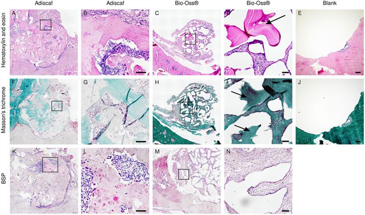 Fig.2 Histological evaluation following in vivo implantation.2
References
- Nakano, Monika, et al. "Enhanced Bone Regeneration by Scaffold-Free Three-Dimensional Constructs of Human Dental Pulp Stem Cells in a Rat Mandibular Defect Model." International Journal of Molecular Sciences (2026). Distributed under Open Access license CC BY 4.0. The image has been modified; only part of the original image. DOI: https://doi.org/10.3390/ijms27020651.
- Cheng, Chen, et al. "Repair of a rat mandibular bone defect by hypertrophic cartilage grafts engineered from human fractionated adipose tissue." Frontiers in Bioengineering and Biotechnology 10 (2022): 841690. Distributed under Open Access license CC BY 4.0. The image has been modified; only part of the original image. DOI: https://doi.org/10.3389/fbioe.2022.841690.
For Research Use Only.