
Arthrofibrosis Modeling & Pharmacodynamics Services
Are you currently facing high attrition rates in orthopaedic drug development, difficulty in replicating human joint stiffness in vivo, or challenges in quantifying fibrotic tissue remodeling? Our Arthrofibrosis Modeling & Pharmacodynamics Services help you accelerate lead optimization and obtain high-resolution pathology data through validated rabbit models and multi-dimensional evaluation platforms. By integrating advanced histomorphometry and biomechanical profiling, Creative Biolabs ensures your therapeutic candidates are tested against the most rigorous standards of joint capsular fibrosis.
Overview of Arthrofibrosis Modeling & Pharmacodynamics Services
Arthrofibrosis is a pathological fibrotic condition characterized by the excessive deposition of extracellular matrix (ECM) within the joint capsule and surrounding soft tissues. This physiological system, primarily involving the synovium and articular capsule, becomes a "biological pressure cooker" following trauma or surgery. Key diseases associated with this condition include post-traumatic joint stiffness, complications following Total Knee Arthroplasty, and "frozen shoulder" (adhesive capsulitis). At its core, the condition is driven by the persistent activation of myofibroblasts, specialized cells expressing α-smooth muscle actin, which contract the joint space. Without intervention, this leads to permanent loss of Range of Motion (ROM), chronic pain, and significant physical disability. Creative Biolabs' services focus on identifying agents that can disrupt this fibrotic cascade at the molecular level.
Arthrofibrosis Models
Creative Biolabs utilizes surgically induced and immobilization-based Rabbit models to replicate the pathophysiology of human joint contracture. Our validated Rabbit and Rat Arthrofibrosis models focus on the knee joint, employing standardized surgical trauma to the femoral condyle combined with external fixation. This approach reliably triggers capsular thickening, synovial hyperplasia, and collagenous adhesion formation within 2 to 8 weeks, providing a robust window for pharmacodynamic intervention.
| Models | Related Disease | Drug Evaluation | Animal Species |
| Arthrofibrosis Models | Acute Achilles tendon rupture, Mid-substance tendinopathy, Paratenonitis, and Haglund's deformity. | Evaluation of pro-regenerative biologics (PRP, BM-MSCs), anti-inflammatory small molecules, collagen-crosslinking agents, and systemic metabolic modulators. | Rat, Rabbit |
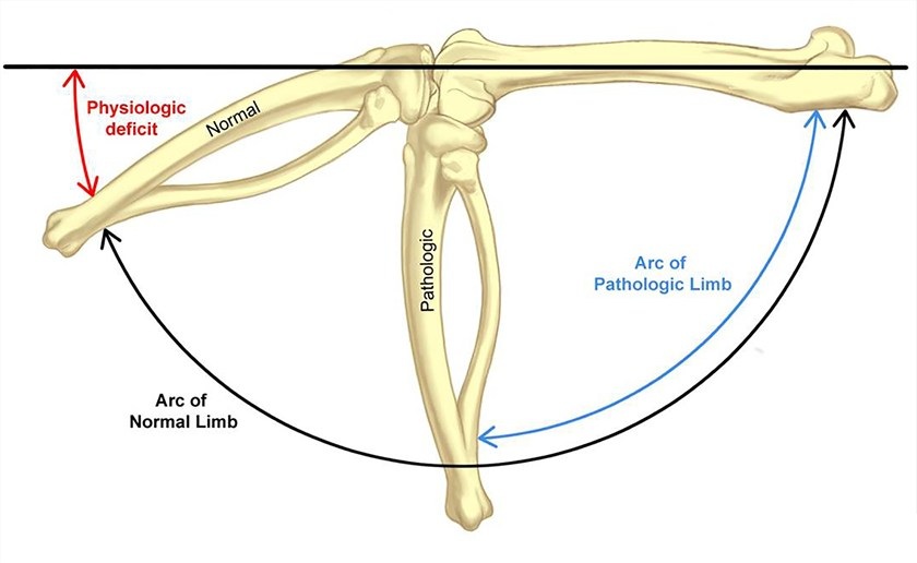 Fig.1 Schema for assessing normal and pathological hind limb conditions in rats.1,3
Evaluation Platform of Our Service
Our platform provides the granular data required to validate therapeutic efficacy at the molecular, cellular, and functional levels.
-
Biochemical & Molecular Analysis:
- Cytokine Profiling (TGF-β1, IL-1β, TNF-α, IL-6) via ELISA
- Hydroxyproline quantification (Total Collagen Content)
- qPCR for fibrotic gene expression
- Western Blot for SMAD signaling pathway activation
-
Histopathological Examination:
- H&E, Masson's Trichrome, and Sirius Red staining
- Capsular thickness measurement and synovial scoring systems
- IHC for myofibroblast markers (α-SMA, Vimentin)
-
Behavioral & Functional Testing:
- Passive Range of Motion (ROM) measurement (Goniometry)
- Joint torque-to-bend analysis (Biomechanical Stiffness)
- Gait analysis and weight-bearing distribution
-
Advanced Imaging & Instrumentation:
- Micro-CT for periarticular bone changes and calcification
- High-resolution MRI for soft tissue fibrosis visualization
- Ultrasonic elastography for capsular elasticity assessment
Key Applications
These models are engineered to simulate clinical indications such as Post-Traumatic Joint Contracture, Frozen Shoulder (Capsulitis), and Post-Surgical Adhesions. Creative Biolabs' evaluation platform is compatible with a wide range of therapeutic modalities:
- Small Molecules: TGF-β inhibitors, anti-inflammatory agents, and kinase inhibitors.
- Biologics: Neutralizing antibodies targeting profibrotic cytokines.
- Gene Therapies: Viral vector-mediated knockdown of collagen-promoting genes.
- Cell-Based Therapies: MSC-derived secretome and exosome treatments for tissue regeneration.
Why Choose Us?
We utilize Rabbit models exclusively for their superior joint size and physiological similarity to human ligamentous structures, ensuring translatable results.
Our workflow covers every stage from initial model induction to complex in vivo PD/PK studies, providing a seamless data package for regulatory filings.
Our PhD-led orthopaedic research team operates under a rigorous quality management system, ensuring reproducibility and scientific integrity in every study.
Work with Us
- Summarize the project requirements and fill in the information collection form.
- Sign a CDA from both parties to further communicate information, such as targets.
- Select an animal model, discuss experimental design, and determine assay parameters.
- Project costing and project schedule forecasting.
- We provide a detailed project plan, including the required sample quantities, methods, and protocols.
- Both parties confirm the project details and start the project.
- Confirm the timeline of the project.
- We provide periodic results and information on the animal's condition.
- We will work together to make project adjustments as necessary.
- We provide a comprehensive project report promptly.
- We arrange transportation for the produced samples.
- We provide a discussion of the project results and help to arrange the next steps.
- Data storage and archiving.
Frequently Asked Questions
-
Q: Why do you prefer Rabbit models over smaller rodents for arthrofibrosis?
A: Rabbits offer a larger joint volume, which is critical for accurate range-of-motion measurements and allows for multiple intra-articular injections and synovial fluid sampling without compromising the model's integrity.
-
Q: Can your platform evaluate the impact of physical therapy combined with drug treatment?
A: Yes, we offer standardized mobilization protocols alongside pharmacological intervention to study synergistic effects on joint recovery.
-
Q: What is the typical duration of an arthrofibrosis pharmacodynamics study?
A: Depending on the model (acute vs. chronic), studies typically range from 4 to 12 weeks, including model induction, treatment window, and terminal analysis.
-
Q: How do you ensure the stability of the fibrotic phenotype?
A: We use standardized surgical trauma and verified immobilization periods, with control groups consistently reaching a predictable "plateau" of joint contracture.
-
Q: Are custom biomarkers available for specific therapeutic targets?
A: Absolutely. Creative Biolabs can develop and validate custom IHC or qPCR assays tailored to your specific molecule's mechanism of action.
Published Data
Objective: To evaluate the efficacy of a next-generation monoclonal antibody in reducing capsular fibrosis by targeting the viral-like immune triggers that drive persistent myofibroblast activation.
Model Used: Surgical trauma-induced Rabbit Knee Arthrofibrosis model with 6 weeks of immobilization.
Results: This model demonstrated high fidelity in replicating clinical capsular stiffness, with therapeutic intervention yielding a 35% increase in passive ROM. Quantitative histology confirmed the model's sensitivity to myofibroblast modulation, showing a significant reduction in α-SMA+ cells and a 40% decrease in hydroxyproline-indexed collagen deposition. By accurately simulating the transition from acute trauma to chronic "Structural Obsolescence," this model provides a robust, clinically relevant platform for validating anti-fibrotic therapies and quantifying the reversal of pathological joint contracture.
Creative Biolabs provides industry-leading Arthrofibrosis Modeling & Pharmacodynamics Services, combining surgical excellence with advanced molecular analytics to de-risk your orthopaedic pipeline. Our specialized focus on Rabbit models ensures that your therapeutic data is both robust and clinically relevant. Contact our specialist team today to discuss your study design and receive a customized proposal.
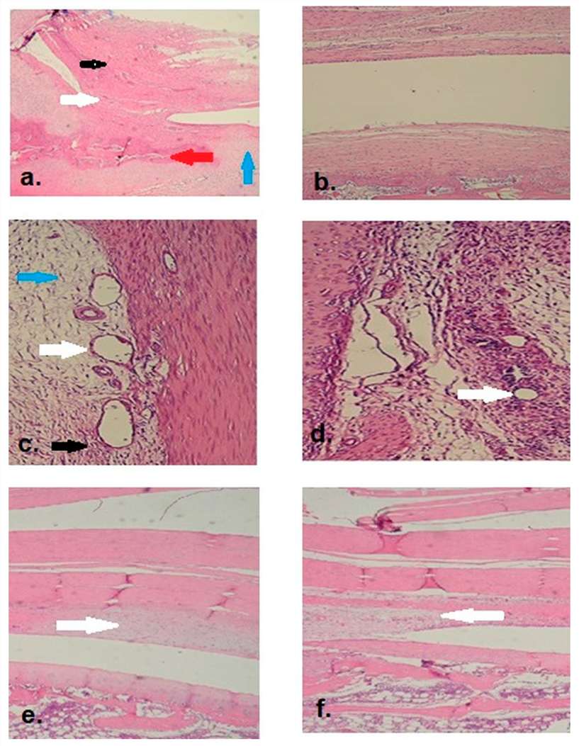 Fig.2 Histological analysis with H&E staining illustrates full knee joint mobility in treated rabbits, contrasted with restricted movement in controls.2,3
References
- Owen, Aaron R., et al. "Biomechanical, histological, and molecular characterization of a new posttraumatic model of arthrofibrosis in rats." Journal of Orthopaedic Research® 40.2 (2022): 323-337. DOI: https://doi.org/10.1002/jor.25054.
- Yagci, Ismail Tugay, et al. "Effect of Carboxymethylcellulose Hyaluronan (SEPRAFİLM®) on an Arthrofibrosis Model Created in Rabbit Knees." Life 15.9 (2025): 1405. DOI: https://doi.org/10.3390/life15091405.
- Distributed under Open Access license CC BY 4.0, without modification.
For Research Use Only.