
Glucocorticoid induced Osteoporosis Modeling & Pharmacodynamics Service
Are you currently facing challenges in assessing the complex osteo-immune mechanisms of bone loss or struggling with inconsistent preclinical models that fail to predict clinical efficacy? Our Glucocorticoid-Induced Osteoporosis (GIOP) Pharmacodynamics Service helps you validate novel therapeutics and obtain high-resolution skeletal data through our optimized rodent platforms and multi-dimensional analytical suites. Creative Biolabs streamlines your R&D process using targeted steroid-induction protocols that mimic human pathophysiology with exceptional fidelity.
Overview of Glucocorticoid-Induced Osteoporosis
Glucocorticoid-Induced Osteoporosis (GIOP) is the most common form of secondary osteoporosis, occurring as a severe adverse effect of long-term glucocorticoid (GC) therapy for inflammatory and autoimmune conditions. Unlike age-related bone loss, GIOP is characterized by a rapid, early-phase decline in bone mineral density (BMD) and a profound disruption of the bone microenvironment. The physiological hallmark of GIOP is a "double-blind" assault on the skeleton: GCs simultaneously trigger the apoptosis of osteoblasts (bone-forming cells) and osteocytes while prolonging the lifespan of osteoclasts (bone-resorbing cells). Recent research highlights that GCs also activate a specific T-cell mediated RANKL surge within the bone marrow, creating an "immuno-skeletal" imbalance. This leads to micro-architectural deterioration and an increased risk of fragility fractures that often precede measurable changes in traditional DXA scans.
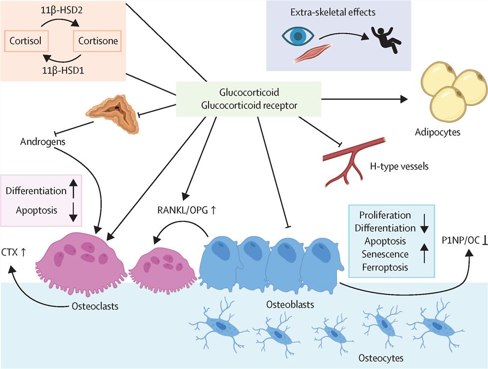 Fig.1 Immediate impact of glucocorticoids on skeletal tissue.1
GIOP Disease Models
Creative Biolabs provides highly validated Rat and Rabbit models designed to simulate different clinical GC exposure scenarios. We utilize precise administration protocols, including subcutaneous pellet implantation, intramuscular injections, or oral dosing of Dexamethasone, Prednisolone, or Methylprednisolone, to induce rapid skeletal catabolism. Our models are optimized to reflect the acute "bone formation gap" and the sarcopenia-osteoporosis synergy frequently observed in patients. These models allow for the evaluation of both preventative strategies and restorative (anabolic) interventions within clinically relevant windows.
Evaluation Platform of Our Service
Creative Biolabs offers a sophisticated analytical suite to capture the granular details of drug efficacy across biochemical, structural, and functional axes.
-
Biochemical & Molecular Analysis
- Bone Turnover Markers: Serum P1NP, CTX-I, Osteocalcin, and BALP.
- Cytokine Profiling: Multiplex assays for RANKL/OPG ratio, TNF-α, IL-6, and Sclerostin levels.
- Molecular Expression: qPCR and Western Blotting for Wnt-signaling pathway components (β-catenin, DKK1).
-
Histopathological Examination
- Bone Histomorphometry: Static and dynamic parameters (MAR, BFR/BS) using fluorescent double-labeling.
- Staining & Scoring: H&E for marrow fat fraction, Goldner's Trichrome for osteoid assessment, and TRAP staining for osteoclast quantification.
- Immunohistochemistry (IHC): Spatial localization of osteocyte apoptosis markers and T-cell infiltration.
-
Behavioral & Functional Testing
- Biomechanical Testing: Three-point bending (long bones) and compression testing (vertebrae) to determine ultimate load and stiffness.
- Motor Coordination: Grip strength and gait analysis to evaluate the muscle-bone unit health.
-
Advanced Imaging & Instrumentation
- Micro-CT Analysis: High-resolution 3D quantification of BV/TV, Tb.N, Tb.Th, and Tb.Sp.
- Bone Mineral Density (BMD): Dual-energy X-ray absorptiometry optimized for small animal precision.
Key Applications
Our GIOP modeling and evaluation services are tailored to simulate a wide range of human indications, including chronic obstructive pulmonary disease, rheumatoid arthritis, and post-transplant immunosuppression-related bone loss. Creative Biolabs supports the evaluation of various therapeutic modalities:
- Small Molecules: Antiresorptives and novel anabolic compounds.
- Biologics: Monoclonal antibodies targeting RANKL or Sclerostin.
- Gene Therapies: Targeted delivery of Wnt-pathway modulators.
- Cell-Based Therapies: Mesenchymal stem cell (MSC) transplantation for bone niche restoration.
Why Choose Us?
We offer meticulously validated Rat and Rabbit strains, allowing for scaling of PK/PD studies and orthopedic surgical modeling.
Our seamless workflow bridges initial in vitro screening with comprehensive in vivo PD/PK studies, providing a unified data package for regulatory submission.
Our PhD-led project teams bring over 20 years of experience in musculoskeletal research, supported by a rigorous quality management system that ensures data reproducibility and scientific integrity.
Work with Us
- Summarize the project requirements and fill in the information collection form.
- Sign a CDA from both parties to further communicate information, such as targets.
- Select an animal model, discuss experimental design, and determine assay parameters.
- Project costing and project schedule forecasting.
- We provide a detailed project plan, including the required sample quantities, methods, and protocols.
- Both parties confirm the project details and start the project.
- Confirm the timeline of the project.
- We provide periodic results and information on the animal's condition.
- We will work together to make project adjustments as necessary.
- We provide a comprehensive project report promptly.
- We arrange transportation for the produced samples.
- We provide a discussion of the project results and help to arrange the next steps.
- Data storage and archiving.
Frequently Asked Questions
-
Q: How quickly can your models show measurable bone loss after GC induction?
A: In our optimized Rat models, significant decreases in bone formation markers and micro-architectural changes are typically observable within 2 to 4 weeks, allowing for rapid-turnaround efficacy testing.
-
Q: Can Creative Biolabs help distinguish between antiresorptive and anabolic drug effects?
A: Absolutely. Our dynamic histomorphometry and Micro-CT services are specifically designed to decouple bone formation from resorption, providing clear evidence of your compound's mechanism of action.
-
Q: What is the benefit of using the Rabbit model over the Rat model for GIOP?
A: Rabbits exhibit Haversian remodeling similar to humans, making them an excellent choice for late-stage preclinical validation and orthopedic device testing where cortical bone structure is critical.
-
Q: How do I initiate a pilot study with Creative Biolabs?
A: We encourage you to reach out for a technical consultation. Our team will help you select the most relevant species and dosing regimen to meet your specific research objectives.
Published Data
Objective: To determine the role of T cells in the pathogenesis of glucocorticoid-induced bone loss and evaluate if targeting the immune-bone axis can mitigate GIOP.
Model Used: Dexamethasone-induced bone loss model in T-cell deficient vs. wild-type rodents (Rat-based validation).
Results: This specialized T-cell-deficient/reconstitution model offers a powerful platform for dissecting immune-skeletal interactions. Data demonstrate that GIOP is significantly attenuated without T cells, identifying bone marrow-resident T cells, which resist GC-induced apoptosis and overexpress RANKL, as the primary drivers of osteoclastogenesis. The model's high sensitivity and phenotypic fidelity were validated by T-cell reintroduction, which restored bone degradation. Consequently, this model serves as a highly translatable tool for screening immunomodulatory agents and evaluating precision therapies targeting the immune-bone axis.
Creative Biolabs provides the scientific depth and technical precision required to navigate the complexities of Glucocorticoid-Induced Osteoporosis. From advanced imaging to molecular profiling in Rat and Rabbit models, we deliver the data you need to move your lead candidates forward with confidence. Our team of experts is standing by to provide a customized project proposal.
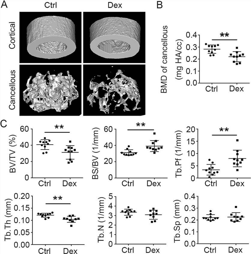 Fig.2 Micro-CT evaluation of femoral bones from wild-type mice with and without drug treatment.2,3
References
- Hofbauer, Lorenz C., et al. "Glucocorticoid-induced osteoporosis: novel concepts and clinical implications." The Lancet Diabetes & Endocrinology 13.11 (2025): 964-979. Distributed under Open Access license CC BY 4.0. DOI: https://doi.org/10.1016/S2213-8587(25)00251-7.
- Song, Lin, et al. "The critical role of T cells in glucocorticoid-induced osteoporosis." Cell death & disease 12.1 (2020): 45. DOI: https://doi.org/10.1038/s41419-020-03249-4.
- Distributed under Open Access license CC BY 4.0. The image has been modified; only part of the original image.
For Research Use Only.