
Retinoic Acid induced Osteoporosis Modeling & Pharmacodynamics Service
Are you currently facing challenges in evaluating bone-targeted therapies due to slow-progressing models or inconsistent bone mineral density data? Our Retinoic Acid-Induced Osteoporosis Modeling & Pharmacodynamics Service helps you obtain rapid, reliable, and high-resolution efficacy data for your lead compounds. Through our specialized retinoic acid induction protocols and multi-dimensional micro-CT imaging, Creative Biolabs accelerates your R&D timeline by providing a high-turnover bone loss environment that mimics acute secondary osteoporosis.
Overview of Retinoic Acid-Induced Osteoporosis
The skeletal system is a dynamic tissue governed by the delicate balance between osteoblast-mediated bone formation and osteoclast-mediated bone resorption. Osteoporosis occurs when this equilibrium shifts toward systemic bone mass reduction and micro-architectural deterioration. Retinoic Acid (RA) induction serves as a potent biochemical trigger that accelerates bone turnover by stimulating osteoclast activity and suppressing osteoblast differentiation. Unlike age-related or hormone-deficiency models, RA-induced models provide a rapid-onset pathology, making them ideal for evaluating the protective effects of small molecules, organic trace elements (like Selenomethionine), and regenerative agents.
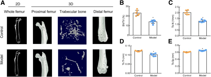 Fig.1 Development of an osteoporosis model induced by retinoic acid.1
Retinoic Acid-Induced Osteoporosis Models
Creative Biolabs offers a robust portfolio of validated rodent models to simulate various clinical manifestations of bone loss. Our Retinoic Acid-Induced Osteoporosis Model is particularly valued for its short induction cycle (typically 2-3 weeks), allowing for high-throughput pharmacodynamic screening. In addition to RA-induction, we provide Orchiectomy models for male-specific bone loss, Ovariectomy for postmenopausal simulations, and Glucocorticoid-Induced models to study secondary osteoporosis. These models are strictly maintained in our AAALAC-accredited facilities using the rat and Rabbit species to ensure physiological relevance.
Evaluation Platform of Our Service
Our platform utilizes high-end instrumentation to quantify bone quality and metabolic markers with surgical precision.
-
Biochemical & Molecular Analysis:
- Bone Turnover Markers: Serum ALP, TRACP-5b, Osteocalcin, and CTX-I.
- Cytokine Profiling: TNF-α, IL-6, and NF-κB pathway analysis via Western Blot.
- Antioxidant Status: GSH-Px activity and ROS quantification.
-
Histopathological Examination:
- Staining: H&E for trabecular structure; Goldner's Trichrome for mineralized bone vs. osteoid.
- Immunohistochemistry (IHC): Expression of Runx2, OCN, and VEGF for angiogenesis coupling.
-
Behavioral & Functional Testing:
- Mechanical Testing: Three-point bending and femoral neck compression tests for bone strength.
- Gait Analysis: Assessing mobility changes in severe osteoporotic Rat models.
-
Advanced Imaging & Instrumentation:
- Micro-CT: 3D quantification of BV/TV, Tb.N, Tb.Th, and Tb.Sp.
- DXA: High-speed Bone Mineral Density (BMD) measurement.
Key Applications
Our Retinoic Acid-Induced Osteoporosis models are designed to simulate acute bone destruction and metabolic mineral imbalance. These models are highly effective for evaluating:
- Targeted Indications: Secondary osteoporosis, metabolic bone disease, and bone loss associated with Vitamin A toxicity.
- Therapeutic Modalities: Small molecule inhibitors, biologics, nutraceuticals (organic selenium, melatonin), and cell-based regenerative therapies targeting Bone Marrow Mesenchymal Stem Cells (BMSCs).
Why Choose Us?
We utilize high-quality Rat and Rabbit strains, ensuring the skeletal architecture is compatible with high-resolution Micro-CT and mechanical testing protocols.
Our workflow bridges the gap between in vitro BMSC differentiation assays and in vivo PD/PK studies, providing a holistic data package for IND submissions.
Led by a PhD-level team with decades of experience in bone biology, Creative Biolabs ensures rigorous quality management and data integrity.
By incorporating the latest research findings, such as the link between positive emotional states and mineral homeostasis, we provide a deeper context for your results.
Work with Us
- Summarize the project requirements and fill in the information collection form.
- Sign a CDA from both parties to further communicate information, such as targets.
- Select an animal model, discuss experimental design, and determine assay parameters.
- Project costing and project schedule forecasting.
- We provide a detailed project plan, including the required sample quantities, methods, and protocols.
- Both parties confirm the project details and start the project.
- Confirm the timeline of the project.
- We provide periodic results and information on the animal's condition.
- We will work together to make project adjustments as necessary.
- We provide a comprehensive project report promptly.
- We arrange transportation for the produced samples.
- We provide a discussion of the project results and help to arrange the next steps.
- Data storage and archiving.
Frequently Asked Questions
-
Q: Can you evaluate both prophylactic and therapeutic effects?
A: Yes. Our protocols allow for "Prevention" (co-administration with RA) and "Treatment" (administration after bone loss is established) phases to define your drug's optimal clinical window.
-
Q: Does Creative Biolabs provide mechanical strength data?
A: Absolutely. We consider bone mineral density only half the story; we perform three-point bending tests to ensure that increases in density translate to actual fracture resistance.
-
Q: Are the Rat and Rabbit models suitable for human-equivalent dosing?
A: Our team provides allometric scaling support to help you translate rodent dosages to clinical targets, utilizing our extensive PK/PD database.
-
Q: How can I start a project with Creative Biolabs?
A: Simply reach out via our inquiry prompt below. We offer a technical consultation to customize the model parameters to your specific molecule's mechanism of action.
Published Data
Objective: To evaluate the protective effects of Melatonin on bone microarchitecture and the osteogenesis-angiogenesis coupling in an osteoporotic environment.
Model Used: Mice (Retinoic Acid-Induced, 2-week induction).
Results: Micro-CT analysis demonstrated that Melatonin significantly reversed RA-induced bone loss, characterized by increased BV/TV and improved trabecular thickness. Mechanistically, the intervention enhanced the osteogenic differentiation of BMSCs and promoted vascularization within the bone marrow, proving the dual-action potential of the therapy.
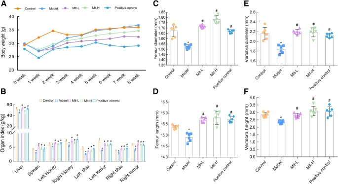 Fig.2 Effects of melatonin on femoral and vertebral (l1) alterations, body weight, and organ index in osteoporotic mice.1
Creative Biolabs provides the biological expertise and high-end technical platform required to transform your bone-health candidates into market-ready therapies. From rapid retinoic acid induction to complex histomorphometry, we are your dedicated partner in bone metabolism research. Contact Our Team for More Information and to Discuss Your Project.
Reference
- Wang, Xudong, et al. "Melatonin prevents bone destruction in mice with retinoic acid-induced osteoporosis." Molecular Medicine 25.1 (2019): 43. Distributed under Open Access license CC BY 4.0. The image has been modified; only part of the original image. DOI: https://doi.org/10.1186/s10020-019-0107-0.
For Research Use Only.