
Meniscectomy (MNX) induced Osteoarthritis Modeling & Pharmacodynamics Service
Are you currently facing challenges in antibody development, long drug development cycles, or complex clinical trials for joint diseases? Our Meniscectomy (MNX)-Induced Osteoarthritis Modeling & Pharmacodynamics Service helps you accelerate drug discovery and streamline clinical trial processes through innovative protein engineering techniques and high-throughput screening platforms. Creative Biolabs provides the precision required to move your lead compounds into the clinic with confidence and speed.
Overview of MNX-Induced Osteoarthritis
Osteoarthritis (OA) is a debilitating degenerative joint disease characterized by the progressive breakdown of articular cartilage, subchondral bone remodeling, and synovial inflammation. The meniscus plays a vital role in shock absorption and load distribution; its removal (meniscectomy) leads to immediate mechanical instability and localized high-stress zones. This physiological trigger induces a rapid onset of OA that mimics the pathology seen in humans following sports injuries or traumatic joint accidents. By focusing on this system, Creative Biolabs allows researchers to study the entire arc of joint failure, from early inflammatory responses to chronic cartilage erosion and osteophyte formation.
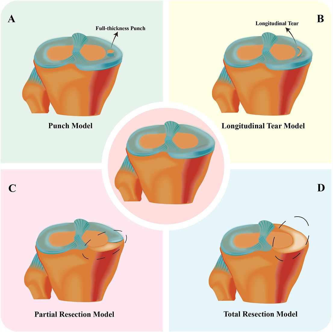 Fig.1 Surgical representation of animal models incorporating four MNX variants.1,3
MNX-Induced Disease Models
Creative Biolabs specializes in highly reproducible surgical models of OA in the rat and Rabbit species. We utilize total or partial medial meniscectomy (MMx) to induce predictable joint degeneration within 4 to 12 weeks post-surgery. These models are characterized by severe fibrillation, loss of proteoglycans, and subchondral bone sclerosis. Our surgical team ensures minimal variability by standardizing incision points and meniscus excision techniques, providing a consistent baseline for evaluating therapeutic efficacy.
Evaluation Platform of Our Service
Our depth of analysis ensures every facet of your drug's performance is captured. Creative Biolabs utilizes the following technical parameters:
-
Biochemical & Molecular Analysis:
- Pro-inflammatory cytokine profiling.
- Catabolic marker detection via Western Blot and qPCR.
- Synovial fluid analysis for hyaluronic acid and lubricant protein concentration.
-
Histopathological Examination:
- Safranin-O/Fast Green and H&E staining.
- Validated scoring systems (OARSI, Mankin, and ICRS scores).
- Immunohistochemistry (IHC) for Type II Collagen and Aggrecan degradation.
-
Behavioral & Functional Testing:
- Gait analysis to measure stride length and paw pressure.
- Nociception assessment for mechanical allodynia.
- Incapacitance testing to measure weight-bearing asymmetry between limbs.
-
Advanced Imaging & Instrumentation:
- High-resolution Micro-CT for subchondral bone mineral density (BMD) and osteophyte volume.
- Contrast-enhanced imaging for volumetric 3D cartilage analysis.
- X-ray radiography for longitudinal joint space measurement.
Key Applications
These models are designed to simulate both post-traumatic and age-related Osteoarthritis. They are highly effective for evaluating:
- Indications: Chronic joint pain, cartilage degradation, subchondral bone remodeling, and synovial inflammation.
- Therapeutic Agents: Small molecule inhibitors, biologics (monoclonal antibodies), gene therapies (AAV-mediated chondrocyte targeting), and cell-based therapies (MSC injections).
Why Choose Us?
Creative Biolabs provides a premium environment for your orthopedic research:
We offer validated, high-health status Rat and Rabbit strains, providing the necessary anatomical scale for diverse delivery methods.
Experience a seamless transition from initial in vitro metabolic flux analysis to definitive in vivo PD/PK studies within a single facility.
Our PhD-level surgical and pathology teams operate under rigorous quality management systems to ensure data integrity and regulatory readiness.
Work with Us
- Summarize the project requirements and fill in the information collection form.
- Sign a CDA from both parties to further communicate information, such as targets.
- Select an animal model, discuss experimental design, and determine assay parameters.
- Project costing and project schedule forecasting.
- We provide a detailed project plan, including the required sample quantities, methods, and protocols.
- Both parties confirm the project details and start the project.
- Confirm the timeline of the project.
- We provide periodic results and information on the animal's condition.
- We will work together to make project adjustments as necessary.
- We provide a comprehensive project report promptly.
- We arrange transportation for the produced samples.
- We provide a discussion of the project results and help to arrange the next steps.
- Data storage and archiving.
Frequently Asked Questions
-
Q: How quickly can we see OA progression in your Rat MNX model?
A: Typically, significant histological changes such as proteoglycan loss appear as early as 2 weeks, with full-thickness cartilage erosion visible by week 4-6, allowing for rapid screening cycles.
-
Q: Is the MNX model superior to the ACLT (Anterior Cruciate Ligament Transection) model?
A: While both are excellent, the MNX model often provides a more rapid and severe induction of cartilage damage, which is particularly useful for testing potent anti-catabolic agents.
-
Q: How do you ensure the pain management of the animals does not interfere with the drug evaluation?
A: We use standardized, multi-modal analgesic protocols that are carefully timed to provide humane care while ensuring no interference with the primary inflammatory markers being measured.
-
Q: What imaging capabilities do you recommend for Rabbit joint studies?
A: For Rabbits, we highly recommend a combination of Micro-CT for bone remodeling and 3D contrast-enhanced imaging to capture volumetric cartilage changes that 2D histology might miss.
Published Data
Objective: To evaluate the efficacy of a continuous therapeutic delivery system on cartilage preservation in a surgical instability model.
Model Used: Surgical instability induced via standardized orthopedic procedures in a high-density environment.
Results: This model precisely simulated the chronic degradative environment of OA, demonstrating that continuous delivery achieved a 40% reduction in Mankin scores and superior Type II Collagen retention via IHC analysis. Unlike traditional models, this platform effectively captured the dose-response relationship of the therapeutic agent under stable physiological concentrations. The results highlight the model's high sensitivity in detecting cartilage matrix preservation, making it a gold-standard vehicle for evaluating long-acting injectables and localized drug delivery systems.
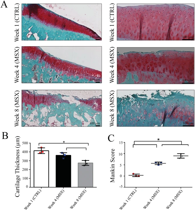 Fig.2 Cartilage sections from medial condyles in control and meniscectomy joints.2,3
Creative Biolabs is your premier partner for Meniscectomy (MNX)-Induced Osteoarthritis Modeling & Pharmacodynamics Service. By combining expert microsurgery, high-resolution 3D imaging, and comprehensive molecular profiling, we provide the clarity your R&D pipeline demands. Our specialist team is standing by to provide detailed protocols and tailored project quotes.
References
- Peng, Xiaoyao, et al. "Current advances in animal model of meniscal injury: From meniscal injury to osteoarthritis." Journal of Orthopaedic Translation (2025). DOI: https://doi.org/10.1016/j.jot.2024.11.005.
- Ali, Tonima S., et al. "Progression of Post-Traumatic Osteoarthritis in rat meniscectomy models: Comprehensive monitoring using MRI." Scientific Reports 8.1 (2018): 6861. DOI: https://doi.org/10.1038/s41598-018-25186-1.
- Distributed under Open Access license CC BY 4.0, without modification.
For Research Use Only.