
Spinal Muscular Atrophy (SMA) Modeling & Pharmacodynamics Service
Introduction
Spinal Muscular Atrophy (SMA) is a devastating autosomal recessive neuromuscular disorder and a leading genetic cause of infant mortality, driven by Survival Motor Neuron 1 (SMN1) gene mutations that lead to survival motor neuron protein deficiency and the progressive degeneration of spinal motor neurons. Creative Biolabs' specialized SMA platform addresses this critical unmet need by bridging advanced genetic engineering with high-fidelity translational analytics, utilizing an extensive library of Global Knockout (KO), Conditional KO, and Knock-In (KI) models to recapitulate disease hallmarks such as muscular atrophy and Neuromuscular Junction (NMJ) dysfunction. By integrating Antisense Oligonucleotides (ASOs) / antisense oligonucleotides (ASOs)-optimized pipelines with rigorous readouts, including Compound Muscle Action Potentials (CMAP) electrophysiology, NMJ morphology, and longitudinal phenotyping, we provide a robust framework to confirm target engagement, optimize delivery routes, and de-risk next-generation therapies across splice-modifying small molecules, oligonucleotides, and viral-mediated gene replacements.
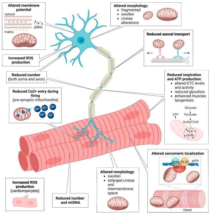 Fig.1 Schematic summary of the mitochondrial defects observed in SMA neurons and SMA muscles.1,3
Available Spinal Muscular Atrophy (SMA) Model
Our platform provides an extensive suite of genetically engineered mouse models, including Global KO, Conditional KO (Flox), and Knock-in (KI) strains, meticulously designed to recapitulate the progressive loss of motor neurons, muscular atrophy, and impaired neuromuscular junction (NMJ) function. By covering a broad spectrum of biological pathways, from ion channel function and protein homeostasis to autophagy and neuroinflammation, these models provide the precise genetic context required to investigate core pathogenic mechanisms and validate next-generation neurotherapeutics. Furthermore, we offer high-specificity Cre driver lines, such as the motor neuron-specific Mnx1-Cre, to enable the precise spatiotemporal gene manipulation essential for advanced mechanistic studies and the rigorous preclinical validation of therapeutic targets for SMA and other neuromuscular disorders.
| SMA Model | Model Types / Key Genes | Research Applications & PD Value | Animal Species |
| Global KO Models | Setx, Grid2, Cnst, IL16, Fgf14, Snx16, Grm1, Pdyn, Kcnc3, Prkcg, Rtn1, Ppp2r2b, Sptbn2, Tdp1, Atp2B3, Zfp592, Cacna1G, Rubcn, Hspa8, Ifrd1, Rrm1, Ccl1, Nop56 | Essential for investigating systemic pathology and identifying early-stage biomarkers across motor neuron degeneration and spinocerebellar ataxias. | Mouse |
| CKO/Flox Models | Ighmbp2, Dync1h1, Cacna1a, Trpc3, Sox9, Carm1, Atp7a, Cnst, Vwc2l, Atp2b3, Setx, Fgf14, Muc1, Pdyn, Zfp592, Kat2a, Afg3l2, Tbp, Xrcc1, Grid2, Cacna1g, Snx16, Atxn7, Eef2, Grm1, Rubcn, IL16, Prkcg, Ppp2r2b, Nop56, Rrm1, Fbln7, Hspa8, Kcnc3, Stub1, Prss8, Ifrd1 | Enables tissue-specific (e.g., motor neuron-specific) or inducible gene deletion to isolate cell-autonomous mechanisms while bypassing embryonic lethality. | Mouse |
| KI Models | Mnx1-IRES-Cre, Eno2-2A-Cre, Krt15-IRES-Cre, Cacna1a-E1980A. | Facilitates precise modeling of clinical point mutations and provides specialized driver lines (e.g., Mnx1, Eno2) for targeted validation of AAV/ASO therapies. | Mouse |
Evaluation Platform
By combining automated motor function assessments (e.g., Rotarod, Grip Strength, Gait Analysis) with deep molecular and electrophysiological profiling, we provide the rigorous, multi-dimensional evidence required to de-risk motor neuron disease candidates from lead optimization through IND-enabling studies.
- Survival & Developmental Milestones: Long-term Longevity Analysis and neonatal Righting Reflex testing to track early-phase developmental recovery and lifespan extension.
- Neuromuscular Integrity & Electrophysiology: Precise functional mapping through CMAP and Motor Unit Number Estimation (MUNE) profiling, alongside high-resolution confocal imaging of NMJ innervation, AChR clustering, and terminal sprouting.
- Quantitative Histopathology: Automated quantification of ChAT-positive alpha-motor neurons in the spinal cord ventral horn and Muscle Fiber Size Distribution (CSA) analysis to measure atrophy rescue.
- Molecular & Genetic Validation: High-sensitivity quantification of SMN protein restoration (ELISA/Wes) and Exon 7 inclusion ratios (RT-qPCR/ddPCR) to validate target engagement and splicing efficiency for AAV/ASO therapies.
Applications
- Mechanistic Research & Lead Optimization: Delivering longitudinal Pharmacodynamics (PD) data through multidimensional profiling of HPA-axis reactivity, neuroinflammation, and synaptic plasticity.
- Gene Therapy Efficacy & Safety Validation: Evaluating AAV9-SMN1 biodistribution, Central Nervous System (CNS) penetrance, and the long-term durability of SMN expression across varied Routes of Administration, such as intrathecal (IT), intracerebroventricular (ICV), and intravenous (IV).
- Synaptic Modulator & Splicing Testing: High-throughput validation of SMN2 Splicing Modifiers (Small Molecules/ASOs) that promote Exon 7 inclusion, and testing of NMDA/GABA modulators for synaptic stabilization.
- Non-SMN Neuroprotective Strategies: Assessing Muscle-Targeted Therapies (e.g., Myostatin inhibitors) and pathways aimed at stabilizing the NMJ and enhancing contractile strength independent of SMN levels.
Our Advantages
- Industry-Leading Genetic Model Library: We offer a sophisticated portfolio of Global KO, CKO/Flox, and KI models. This includes specialized driver lines like Mnx1-Cre (motor neuron-specific) and Eno2-Cre (pan-neuronal), providing the precise genetic context for both systemic and tissue-specific therapeutic validation.
- Gold-Standard "Synapse-to-Symptom" Analytics: Our platform bridges molecular target engagement with functional outcomes. We correlate SMN protein restoration (ELISA/Wes) and Exon 7 splicing ratios (ddPCR) with high-fidelity CMAP/MUNE electrophysiology and NMJ structural integrity.
- High-Resolution Neuromuscular Mapping: We utilize advanced confocal microscopy to quantify AChR clustering, terminal sprouting, and innervation status. This is paired with automated alpha-motor neuron counting (ChAT+) and muscle fiber Cross-Sectional Area (CSA) analysis to provide definitive proof of neuroprotection and atrophy rescue.
- AAV & ASO Optimized Workflows: Specifically designed for modern modalities, our workflows include comprehensive Biodistribution studies and CNS Penetrance analysis. We offer specialized Routes of Administration (IT, ICV, and IV) to optimize delivery and evaluate the durability of gene expression or splicing correction.
- Translational Motor Phenotyping: To ensure regulatory-grade data, we employ a rigorous battery of developmental and motor tests, including the neonatal Righting Reflex, automated Gait Analysis (DigiGait), and Grip Strength testing, capturing the full spectrum of disease progression and therapeutic recovery.
Work with Us
- Summarize the project requirements and fill in the information collection form.
- Sign a CDA from both parties to further communicate information, such as targets.
- Select an animal model, discuss experimental design, and determine assay parameters.
- Project costing and project schedule forecasting.
- We provide a detailed project plan, including the required sample quantities, methods, and protocols.
- Both parties confirm the project details and start the project.
- Confirm the timeline of the project.
- We provide periodic results and information on the animal's condition.
- We will work together to make project adjustments as necessary.
- We provide a comprehensive project report promptly.
- We arrange transportation for the produced samples.
- We provide a discussion of the project results and help to arrange the next steps.
- Data storage and archiving.
FAQs
-
Q: Can you perform IT injections in neonatal mice?
A: Yes, we have specialized micro-injection protocols for neonatal IT delivery to bypass the Blood-Brain Barrier for ASO or viral vector testing.
-
Q: Is it possible to study muscle-specific effects without affecting the spinal cord?
A: Yes, we can utilize local intramuscular (IM) injections or skeletal muscle-specific conditional Smn models.
-
Q: How do I select the optimal model for my drug (ASO vs. AAV)?
A: For ASOs, we recommend models carrying the human SMN2 transgene (e.g., Delta7) to directly measure Exon 7 splicing correction and protein restoration. For AAVs, we focus on models with a clear therapeutic window to quantify SMN1 transgene expression durability and long-term phenotypic rescue.
-
Q: How do you validate that the drug reached the target motor neurons?
A: We utilize ddPCR for vector copy number (VCN) in the spinal cord, combined with Immunofluorescence (IF) to confirm drug/protein localization within ChAT-positive motor neurons.
-
Q: Is behavioral improvement enough to prove functional recovery?
A: While the righting reflex is a key behavioral marker, we use CMAP and MUNE profiling to provide definitive evidence of functional nerve-muscle connectivity and motor unit preservation.
-
Q: What are the most sensitive early-stage markers for efficacy?
A: Before gross symptoms improve, stabilization of the NMJ, specifically reduced denervation, improved AChR clustering, and reduced terminal sprouting, serves as the most sensitive early readout.
-
Q: Can you compare different Routes of Administration?
A: We support head-to-head comparisons of IT, ICV, and IV delivery. We analyze CNS Penetrance and systemic biodistribution to optimize dosing and minimize off-target toxicity (e.g., liver accumulation).
-
Q: How do you study genes that cause embryonic lethality?
A: We use Mnx1-Cre or other driver lines to delete genes in specific tissues. This bypasses developmental death and allows for the study of cell-autonomous vs. non-cell-autonomous drug effects.
Published Data
Neonatal ICV injection of AAV9-coSMN1 in SMA3 mice provides dose-dependent phenotypic rescue (tail retention) and increases lumbar ChAT+ motor neuron density. The efficacy exhibits a plateau effect, indicating a dose saturation point where higher concentrations provide no additional therapeutic advantage.
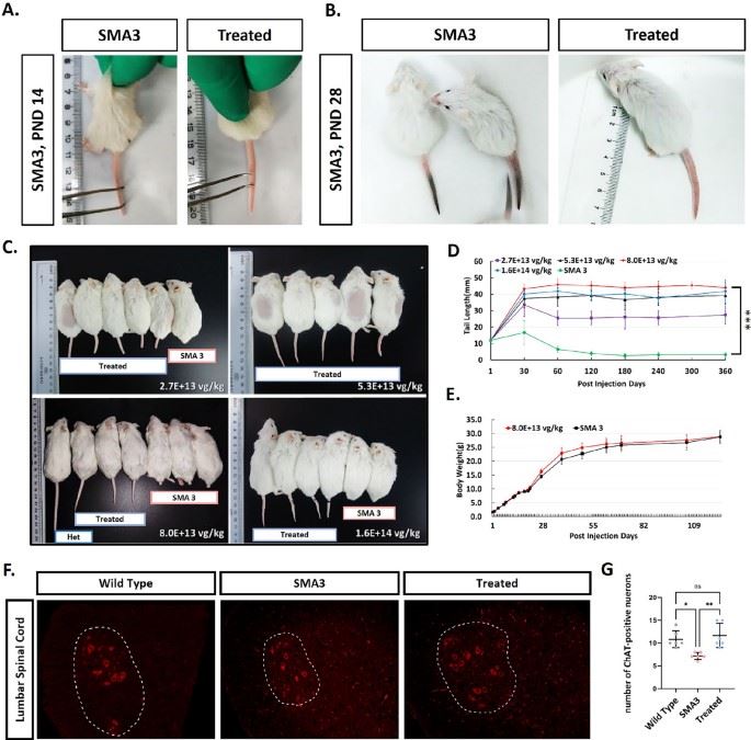 Fig. 2 Motor neurons rescued and tail retention following AAV9-coSMN1 treatment one year.2,3
References
- Zilio, Eleonora et al. "Mitochondrial Dysfunction in Spinal Muscular Atrophy." International Journal of Molecular Sciences vol. 23,18 10878. https://doi.org/10.3390/ijms231810878
- Ma, Wenhao et al. "Preclinical evaluation of AAV9-coSMN1 gene therapy for spinal muscular atrophy: efficacy and safety in mouse models and non-human primates." Molecular Medicine (Cambridge, Mass.) vol. 31,1 158. https://doi.org/10.1186/s10020-025-01207-4
- Distributed under Open Access license CC BY 4.0, without modification.
For Research Use Only.