
Medial Meniscal Tear (MMT) induced Osteoarthritis Modeling & Pharmacodynamics Service
Are you currently facing challenges in assessing DMOAD efficacy, inconsistent disease progression in surgical models, or difficulty correlating structural repair with functional pain relief? Our Medial Meniscal Tear (MMT)-Induced Osteoarthritis Modeling & Pharmacodynamics Service helps you obtain high-fidelity preclinical data and accelerate drug discovery through standardized microsurgical techniques and multi-dimensional longitudinal analysis platforms. We provide the biological complexity needed to move your candidate from bench to clinic with confidence.
Overview of MMT-Induced Osteoarthritis Modeling
The Medial Meniscal Tear (MMT) model is a premier surgical platform used to simulate post-traumatic osteoarthritis (PTOA). The knee joint is a complex physiological system where the meniscus acts as a vital shock absorber, distributing mechanical loads across the articular cartilage. In the MMT model, a precise transection of the medial meniscus is performed, leading to joint instability and altered biomechanics. This instability triggers a cascade of degenerative events: proteoglycan loss, chondrocyte apoptosis, and eventual full-thickness cartilage erosion. Unlike chemical models that cause immediate metabolic cell death, the MMT model replicates the slow, progressive nature of human OA, making it the "gold standard" for evaluating Disease-Modifying Osteoarthritis Drugs (DMOADs) that aim to protect joint structure or alleviate chronic mechanical pain.
MMT-Induced Osteoarthritis Models
Creative Biolabs offers highly standardized surgical MMT models in Rats and Rabbits. Our protocols focus on creating reproducible joint instability to ensure consistent OA progression. By utilizing these rodent and lagomorph species, we provide researchers with the flexibility to perform high-throughput screening or detailed physiological studies requiring larger tissue volumes for biochemical analysis.
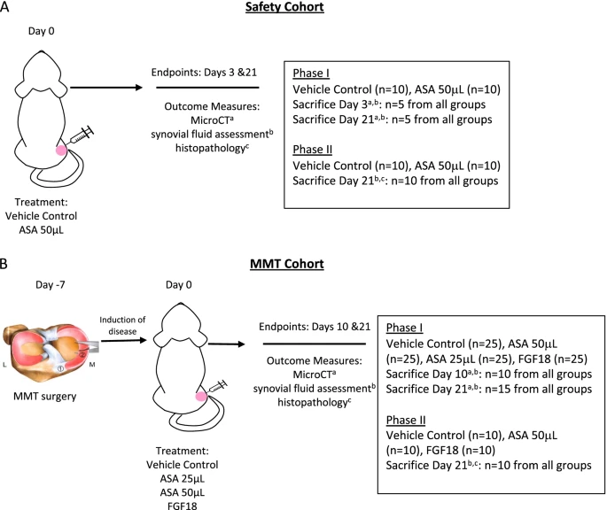 Fig.1 Schema of the study framework for both the safety cohort and the MMT surgical cohort.1,3
Evaluation Platform of Our Service
To ensure your therapeutic candidate is evaluated with the highest scientific rigor, Creative Biolabs has established a comprehensive analytical suite.
-
Biochemical & Molecular Analysis:
- Cytokine Assays: Multiplex ELISA for IL-1β, TNF-α, and IL-6 in synovial fluid.
- Degradation Markers: Analysis of collagen type II and COMP (cartilage oligomeric matrix protein).
- Western Blot/qPCR: Evaluation of MMP-13, ADAMTS-5, and Aggrecan expression levels.
-
Histopathological Examination:
- Tissue Staining: Safranin-O/Fast Green and Toluidine Blue for proteoglycan density.
- Scoring Systems: Standardized OARSI and Modified Mankin scoring by board-certified pathologists.
- IHC: Immunohistochemical localization of Collagen II and inflammatory markers.
-
Behavioral & Functional Testing:
- Incapacitance Testing: Dual-channel weight-bearing asymmetry measurement.
- Gait Analysis: Automated digital footprint tracking to assess stride length and pressure.
- Mechanical Allodynia: Von Frey filament testing for secondary hyperalgesia.
-
Advanced Imaging & Instrumentation:
- Micro-CT (EPIC-µCT): 3D quantitative analysis of cartilage volume, thickness, and surface roughness.
- Subchondral Bone Analysis: Measurement of bone mineral density (BMD), BV/TV, and trabecular thickness.
- Osteophyte Assessment: 3D visualization and volume quantification of mineralized and cartilaginous outgrowths.
Key Applications
Our MMT-induced modeling services are designed to simulate various clinical indications and therapeutic modalities:
- Indications: Post-traumatic osteoarthritis (PTOA), chronic meniscus injury-related degeneration, and subchondral bone sclerosis.
- Therapeutic Agents: Small Molecules: Orally active inhibitors of degradative enzymes.
- Biologics: Intra-articular monoclonal antibodies or growth factors.
- Gene Therapies: Viral vector-mediated chondrocyte reprogramming.
- Cell-Based Therapies: MSC-based regenerative medicine applications.
Why Choose Us?
We specialize exclusively in Rat and Rabbit models, providing deep validation data for these specific translational pathways.
Our workflow covers everything from initial surgical induction to long-term (12-week) longitudinal pharmacodynamic studies, including PK/PD correlations.
Your project is managed by a PhD-level team with over two decades of experience in orthopedic surgery and pathology, ensuring data integrity under rigorous quality management.
Work with Us
- Summarize the project requirements and fill in the information collection form.
- Sign a CDA from both parties to further communicate information, such as targets.
- Select an animal model, discuss experimental design, and determine assay parameters.
- Project costing and project schedule forecasting.
- We provide a detailed project plan, including the required sample quantities, methods, and protocols.
- Both parties confirm the project details and start the project.
- Confirm the timeline of the project.
- We provide periodic results and information on the animal's condition.
- We will work together to make project adjustments as necessary.
- We provide a comprehensive project report promptly.
- We arrange transportation for the produced samples.
- We provide a discussion of the project results and help to arrange the next steps.
- Data storage and archiving.
Frequently Asked Questions
-
Q: Why choose the MMT surgical model over chemical models?
A: Chemical models induce rapid cell death, which doesn't mimic human disease etiology. The MMT model provides the biomechanical instability necessary to test if your drug can actually protect the joint under mechanical stress.
-
Q: What is the typical study duration for an MMT project?
A: While early changes appear at 3 weeks, we recommend 6-12-week studies for a comprehensive look at subchondral bone remodeling and established disease treatment.
-
Q: Can you evaluate pain and structural changes in the same animal?
A: Yes! Our integrated platform allows for longitudinal gait analysis and weight-bearing tests, followed by terminal histopathology and Micro-CT.
-
Q: How do you ensure surgical consistency across large cohorts?
A: All surgeries are performed by a dedicated microsurgery team using standardized SOPs and magnification, minimizing variability between animals.
Published Data
Objective: Characterize the longitudinal progression of OA and the therapeutic window for intervention in the rat MMT model.
Model Used: Rat Medial Meniscus Transection (MMT).
Results: EPIC-μCT imaging revealed a highly reproducible, three-stage progression: Early-stage (3w), marked by proteoglycan depletion and compensatory swelling; Mid-stage (6w), featuring osteophyte formation and subchondral plate thickening; and Late-stage (12w), characterized by full-thickness chondral erosion. Notably, the model's high sensitivity demonstrated that bone-modifying agents were only efficacious during the early active resorption phase, validating the MMT model as a superior platform for stage-specific drug screening and for identifying critical "point-of-no-return" transitions in OA pathology.
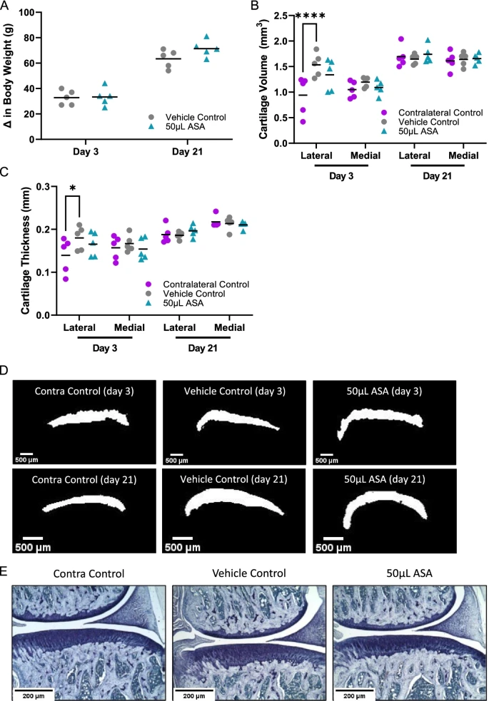 Fig.2 Cartilage and body weight change with Micro-CT and histopathology analyses.1,3
Creative Biolabs provides a world-class platform for MMT-induced Osteoarthritis modeling, combining surgical precision with advanced 3D imaging and functional pain assessment. Whether you are developing a preventative chondroprotector or a regenerative cell therapy, our specialized Rat and Rabbit models offer the translational power needed to advance your program. Our team is standing by to provide a customized study design and quote.
References
- Kimmerling, Kelly A., et al. "Amniotic suspension allograft improves pain and function in a rat meniscal tear-induced osteoarthritis model." Arthritis Research & Therapy 24.1 (2022): 63. DOI: https://doi.org/10.1186/s13075-022-02750-9.
- Qian, Jia-jia, et al. "Expression of VEGF-A signaling pathway in cartilage of ACLT-induced osteoarthritis mouse model." Journal of orthopaedic surgery and research 16.1 (2021): 379. DOI: https://doi.org/10.1186/s13018-021-02528-w.
- Distributed under Open Access license CC BY 4.0, without modification.
For Research Use Only.