
Osteonecrosis of the Femoral Head (ONFH) Modeling & Pharmacodynamics Services
Are you currently facing high clinical failure rates due to animal models that fail to simulate the mechanical collapse of the femoral head? Our ONFH Modeling & Pharmacodynamics Services help you obtain highly predictive preclinical data and accelerate your regulatory submissions through innovative 3D-printed biomechanical induction and genetically enhanced MSC therapies. Creative Biolabs streamlines your R&D by bridging the gap between ischemic pathology and structural restoration.
Overview of ONFH Modeling & Pharmacodynamics Services
Osteonecrosis of the Femoral Head (ONFH) is a debilitating pathological process characterized by the death of osteocytes and bone marrow components due to an interrupted blood supply. This ischemia initiates a cascade of subchondral bone resorption and microfractures, eventually leading to the structural collapse of the weight-bearing portion of the femoral head. While the disease can be traumatic (resulting from neck fractures) or non-traumatic (induced by glucocorticoids or alcohol), the clinical endpoint, severe pain and hip joint disability, is consistent. At Creative Biolabs, we specialize in simulating this complex environment, focusing on the critical window where therapeutic intervention can prevent permanent deformity.
Advanced ONFH Disease Models
Creative Biolabs offers a robust portfolio of validated ONFH models tailored to specific R&D needs. We provide Traumatic Models in Rabbits via surgical vascular deprivation and Non-Traumatic Models in Rats and Rabbits using optimized Glucocorticoid (GC) + LPS induction protocols. Our proprietary Biomechanical-Integrated Models utilize 3D-printed compression devices to simulate the physiological stress concentration that triggers femoral head collapse. Additionally, we offer Transgenic Cell-Based Models to evaluate the synergistic effects of gene therapy and stem cell transplantation in an ischemic microenvironment.
| Models | Related Disease | Drug Evaluation | Animal Species |
| Osteonecrosis of the Femoral Head (ONFH) models | Covers Glucocorticoid-induced metabolic necrosis, Traumatic ischemia (fracture-related), and Alcohol-induced adipogenesis. Models also simulate Idiopathic ONFH and subchondral collapse, capturing clinical ARCO Stage I-IV progression and ischemic bone marrow failure. | Evaluates Regenerative Biologics (VEGF/HGF), Cell Therapies (Transgenic MSCs/Exosomes), and Small Molecules for vascular repair. Also validates Medical Devices, including 3D-printed scaffolds, bioactive grafts, and core decompression tools for structural restoration. | Rat, Rabbit |
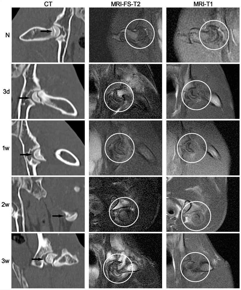 Fig.1 Standard CT and MR imaging of the traumatic ONFH rabbit model.1
Evaluation Platform of Our Service
Our technical platform delivers high-resolution data across four critical analytical dimensions to ensure a comprehensive understanding of drug efficacy.
-
Biochemical & Molecular Analysis:
- Pro-angiogenic Profiling: VEGF, HGF, and bFGF expression levels via ELISA and qPCR.
- Signaling Pathway Analysis: Western Blot for p-ERK1/2, p-Akt, and MAPK activation.
- Cellular Proliferation: PCNA and Ki-67 immunohistochemical quantification.
-
Histopathological Examination:
- Bone Vitality Scoring: Automated quantification of empty bone lacunae ratios in the subchondral zone.
- Tissue Architecture: H&E, Masson's Trichrome, and Safranin O/Fast Green staining for cartilage integrity.
- Bone Formation Markers: Immunohistochemistry (IHC) for Osteocalcin (OCN) and Collagen Type I/II.
-
Behavioral & Functional Testing:
- Gait Analysis: Kinetic tracking of weight-bearing symmetry and stride length.
- Motor Coordination: Modified rotarod testing for assessing joint function in rodents.
- Static Weight Bearing: Measurement of postural changes induced by hip pain.
-
Advanced Imaging & Instrumentation:
- Micro-CT Reconstruction: High-resolution BV/TV, Trabecular Thickness (Tb.Th), and Trabecular Number (Tb.N).
- Bone Mineral Density (BMD): DEXA scanning for longitudinal skeletal assessment.
- Vascular Perfusion: Micro-angiography to visualize microvessel density (MVD) in the femoral neck.
Key Applications
Our platforms are designed to simulate clinical ARCO Phase I through Phase IV progression. Creative Biolabs' models are ideal for evaluating a wide range of therapeutic modalities:
- Small Molecules: Ischemia-reversing agents and anti-inflammatory compounds.
- Biologics: Growth factors (HGF, VEGF) and monoclonal antibodies targeting bone resorption.
- Gene Therapies: Transgenic vectors (e.g., AAV, Lentivirus) for localized protein expression.
- Cell-Based Therapies: Autologous or allogeneic MSCs, EPCs, and exosome-based treatments.
- Medical Devices: Bioactive scaffolds, 3D-printed bone grafts, and core decompression tools.
Why Choose Us?
We provide highly validated strains of Rat and Rabbit, ensuring the optimal balance between cost-efficiency and human-like anatomical fidelity.
Creative Biolabs offers a seamless transition from in vitro BMSC modeling and genomic modification to in vivo PD/PK studies and terminal histopathological validation.
Our projects are managed by experts with decades of experience in orthopedic pathology, ensuring rigorous quality management and peer-review-ready data.
By integrating 3D-printed biomechanics, we offer higher predictive value than traditional chemical-only models.
Work with Us
- Summarize the project requirements and fill in the information collection form.
- Sign a CDA from both parties to further communicate information, such as targets.
- Select an animal model, discuss experimental design, and determine assay parameters.
- Project costing and project schedule forecasting.
- We provide a detailed project plan, including the required sample quantities, methods, and protocols.
- Both parties confirm the project details and start the project.
- Confirm the timeline of the project.
- We provide periodic results and information on the animal's condition.
- We will work together to make project adjustments as necessary.
- We provide a comprehensive project report promptly.
- We arrange transportation for the produced samples.
- We provide a discussion of the project results and help to arrange the next steps.
- Data storage and archiving.
Frequently Asked Questions
-
Q: What is the typical success rate for your non-traumatic ONFH models?
A: Using our optimized GC + LPS protocols, we consistently achieve induction success rates exceeding 70% in rabbits, with clear markers of bone marrow necrosis and trabecular thinning within 4-8 weeks.
-
Q: Do you offer customized transgenic stem cell services for ONFH research?
A: Absolutely. We can engineer MSCs to overexpress specific growth factors (such as HGF) to evaluate their impact on endogenous repair peak windows.
-
Q: How do you ensure the stability of the mechanical stress in your 3D-printed models?
A: We use high-precision resin materials, ensuring the compression device remains structurally stable throughout the 8-week study duration.
-
Q: Can we monitor disease progression longitudinally without euthanizing the animals?
A: Yes, we utilize in vivo Micro-CT and bone mineral density (BMD) measurements at specific intervals to track the efficacy of your treatment over time.
Published Data
Objective: To evaluate the therapeutic efficacy of HGF-transgenic Mesenchymal Stem Cells (MSCs) in accelerating endogenous repair during the acute phase of traumatic ONFH.
Model Used: Rabbit Traumatic ONFH Model (Severed femoral neck and annular ligament).
Results: Micro-CT and IHC analysis revealed that HGF-transgenic MSCs significantly reduced the empty lacunae ratio and increased CD105+ microvessel density. The treatment captured the "2-week repair peak," resulting in significantly higher BV/TV ratios and improved mechanical stability compared to standard MSC groups. This data demonstrates that genetic enhancement of cell therapies is vital for overcoming ischemic thresholds in ONFH.
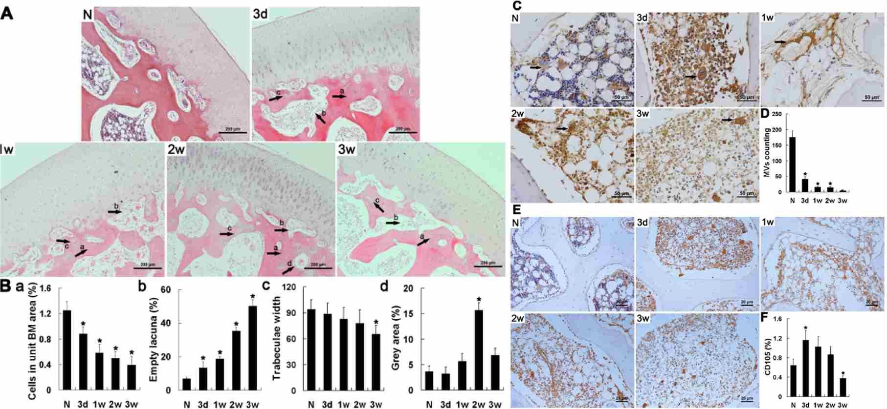 Fig.2 Histopathology of the traumatic ONFH rabbit model, accompanied by immunohistochemical staining and semi-quantitative assessment.1
Creative Biolabs provides the precision modeling and analytical depth required to transform experimental orthopedic concepts into clinical realities. Our integrated approach ensures that your therapeutic candidates are tested against the most rigorous physiological standards. Contact Our Team for More Information and to Discuss Your Project. Our specialists are ready to provide detailed protocols and customized quotes tailored to your specific research goals.
Reference
- Wen, Qian, et al. "HGF-transgenic MSCs can improve the effects of tissue self-repair in a rabbit model of traumatic osteonecrosis of the femoral head." PloS one 7.5 (2012): e37503. Distributed under Open Access license CC BY 4.0, without modification. DOI: https://doi.org/10.1371/journal.pone.0037503.
For Research Use Only.