
Stroke & Brain Ischemia Modeling & Pharmacodynamics Services
Introduction
Stroke remains a leading cause of global adult disability and mortality, with ischemic stroke accounting for approximately 87% of all cases. The complex pathophysiology of stroke involves an immediate "ischemic core" of necrotic tissue surrounded by a salvageable "ischemic penumbra." Successful drug development necessitates robust preclinical models that accurately simulate vessel occlusion, reperfusion injury, and the subsequent neuroinflammatory cascade. To address these challenges, Creative Biolabs provides a sophisticated Stroke & Brain Ischemia research platform centered on clinical translatability. By integrating high-precision surgical models, ranging from focal MCAO and Photothrombotic ischemia to Global Ischemia (4-VO), with advanced blood-brain barrier (BBB) integrity analysis, we empower partners to accelerate the development of next-generation therapeutics. Our integrated approach delivers the high-quality, IND-enabling data required to bridge the gap between benchside discovery and bedside intervention.
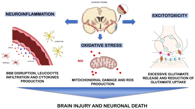 Fig.1 A summary of the pathophysiology involved in ischemic stroke.1
Available Stroke & Brain Ischemia Models
We offer a diverse portfolio of focal and global ischemia models meticulously designed to align with specific therapeutic objectives, ranging from acute neuroprotection to chronic functional rehabilitation. Building upon our established expertise in tMCAO (Transient Middle Cerebral Artery Occlusion) Models, pMCAO (Permanent Middle Cerebral Artery Occlusion) Models, Photochemically induced Ischemic Stroke Model, Collagenase induced Hemorrhagic Stroke Model, and Sodium Laurate induced Cerebral Microvascular Injury Model, Non-human Primates (NHPs) Cerebral Infarction Model, NHPs Vascular Dementia (VaD) Models, and NHPs Cerebral Stroke Model, we have expanded our portfolio to include the following models, providing broader options and greater flexibility for preclinical pharmacodynamics (PD) research and drug development.
| Stroke & Brain Ischemia Models | Modeling Methods | Application Values | Animal Species |
| Endothelin-1 Rodent Model of Stroke & Brain Ischemia | Local microinjection of ET-1 (a potent vasoconstrictor) into specific brain regions (e.g., striatum or cortex). | This model creates targeted, reproducible ischemic lesions with high spatial precision, making it ideal for studying lacunar strokes and localized white matter injury. It is frequently used to evaluate vasodilators and calcium channel blockers, such as Nimodipine, which target vascular constriction. | Rat, Mouse |
| Rodent Photothrombotic Ischemia Model | Systemic injection of Rose Bengal dye followed by focal laser irradiation through the skull. | By inducing precise micro-thrombosis with minimal surgical trauma, this model is the premier choice for research into neuroplasticity, functional brain mapping, and the efficacy of anti-thrombotic agents. It is widely utilized to test thrombolytics like t-PA and various novel anti-platelet therapies. | Mouse, Rat |
| Global Ischemic 4-VO Rat Model | Permanent occlusion of both vertebral arteries combined with transient occlusion of both common carotid arteries. | This model simulates clinical cardiac arrest or severe global hypoperfusion, specifically targeting the vulnerability of hippocampal CA1 neurons. It serves as a robust platform for testing neuroprotectants that combat excitotoxicity and oxidative stress, such as Edaravone or Riluzole. | Rat |
Evaluation Platform
Our platform provides a multi-dimensional assessment of stroke progression and therapeutic efficacy.
-
Neuroimaging & Blood Flow:
- Laser Doppler/Speckle Imaging: Real-time monitoring of cerebral blood flow (CBF) to confirm successful occlusion and reperfusion.
- Small Animal MRI (T2/DWI): Non-invasive quantification of infarct volume and edema at multiple time points.
-
Histopathology & Infarct Analysis:
- TTC Staining: Rapid, reliable quantification of fresh infarct volume within 24–72 hours.
- Nissl & H&E Staining: Assessment of neuronal loss and tissue architecture in chronic phases.
- IHC for Neuroinflammation: Tracking microglial (Iba1) and astrocyte (GFAP) responses in the peri-infarct zone.
-
Neurological & Behavioral Scoring:
- Longa/mNSS Score: Standardized 5- or 18-point scales for acute neurological deficits.
- Cylinder & Grid Walk Tests: Sensitive measures of forelimb asymmetry and fine motor coordination.
- Morris Water Maze: Evaluating cognitive deficits following global ischemia or large cortical strokes.
Applications
- Thrombolytic & Antithrombotic Research: Validating novel "clot-busting" agents, anticoagulants, and mechanical thrombectomy adjuncts in MCAO and embolic models to restore blood flow while minimizing hemorrhagic transformation.
- Acute Neuroprotective Screening: Evaluating molecules that inhibit the ischemic cascade, including excitotoxicity, oxidative stress, and acid-toxicity (ASICs), to preserve neuronal viability during the critical reperfusion window.
- Neuroinflammation & Barrier Integrity: Quantifying BBB penetration (Kp/Kuu) and assessing the effects of modulators on systemic immune cell infiltration, tight junction stability, and post-stroke edema/glial scarring.
- Neural Repair & Plasticity: Validating cell therapies (MSCs/NSCs) and growth factors that promote axonal sprouting, synaptogenesis, and functional circuit rewiring during the chronic recovery phase.
- Translational Functional Recovery: Correlating MRI-based infarct volume reduction with long-term motor and cognitive improvements using a standardized battery of translational neurobehavioral assessments.
- Preclinical Drug Development: Accelerating lead optimization through comprehensive PK/PD profiling and robust IND-enabling efficacy studies to drive rapid advancement of next-generation stroke therapies.
Our Advantages
- Precision ET-1 Micro-Ischemia: Utilizing stereotaxic microinjection of Endothelin-1, we create lacunar strokes with high spatial resolution and zero surgical trauma to the skull or vasculature. This refined platform is ideal for investigating white matter integrity and small vessel disease (SVD) in a highly controlled environment.
- Targeted Photothrombotic Cortical Lesions: Our laser-induced thrombosis can be mapped to precise functional coordinates (e.g., the primary motor cortex). With the industry's lowest mortality rates and most consistent lesion volumes, this model is the premier choice for longitudinal neuroplasticity and functional recovery studies.
- Robust 4-VO Global Ischemia: Our refined 4-Vessel Occlusion (4-VO) technique ensures consistent, bilateral hippocampal CA1 pyramidal cell loss. It serves as a rigorous platform for validating neuroprotectants against cardiac arrest-induced brain injury and associated cognitive decline.
- Real-Time CBF Monitoring (LSCI): We utilize Laser Speckle Contrast Imaging (LSCI) to provide full-field, high-resolution maps of Cerebral Blood Flow (CBF). This real-time validation ensures that every ET-1 or Photothrombotic induction crosses the ischemic threshold before the study proceeds.
- 7T Small Animal MRI: Our facility features high-field MRI for longitudinal tracking. We employ Diffusion-Weighted Imaging (DWI) to visualize acute cytotoxic edema and Perfusion-Weighted Imaging (PWI) to identify the "Ischemic Penumbra", the critical therapeutic window for neuroprotective candidates.
- Ultra-Sensitive Biomarker Quantification: Leveraging Simoa and MSD platforms, we detect low-abundance markers such as Neurofilament Light (NfL) and UCH-L1 in plasma or CSF, providing a high-sensitivity molecular window into axonal damage and disease progression.
- Automated Kinematic Behavioral Analysis: Moving beyond subjective scoring, our DigiGait and CatWalk systems quantify over 50 gait parameters. This objective approach detects subtle motor improvements and subtle pharmacological effects that traditional manual scales (e.g., mNSS) might overlook.
- AI-Driven Quantitative Histopathology: We combine automated slide scanning with AI-driven analysis to quantify neuronal survival (NeuN), synaptic density (Synaptophysin), and glial scarring (GFAP/Iba1) across whole-brain sections, ensuring unbiased and statistically robust data.
Work with Us
- Summarize the project requirements and fill in the information collection form.
- Sign a CDA from both parties to further communicate information, such as targets.
- Select an animal model, discuss experimental design, and determine assay parameters.
- Project costing and project schedule forecasting.
- We provide a detailed project plan, including the required sample quantities, methods, and protocols.
- Both parties confirm the project details and start the project.
- Confirm the timeline of the project.
- We provide periodic results and information on the animal's condition.
- We will work together to make project adjustments as necessary.
- We provide a comprehensive project report promptly.
- We arrange transportation for the produced samples.
- We provide a discussion of the project results and help to arrange the next steps.
- Data storage and archiving.
FAQs
-
Q: How do you distinguish between "brain edema" and "actual infarct tissue"?
A: In the acute phase (24–72 hours), brain swelling (edema) can artificially inflate infarct volume measurements. We use Edema Correction Formulas during TTC or MRI analysis to calculate the "indirect infarct volume," which subtracts the volume of the swelling from the total hemispheric volume to provide a true measurement of tissue death.
-
Q: How is the infarct volume quantified?
A: For rapid screening, we use TTC (2,3,5-triphenyltetrazolium chloride) staining, which identifies metabolically active tissue (red) vs. infarcted tissue (white). For longitudinal studies in the same animal, we use 7T Small Animal MRI (T2-weighted and DWI sequences).
-
Q: What biomarkers do you monitor during stroke studies?
A: We frequently monitor Neurofilament Light (NfL) and S100B in the plasma as markers of axonal damage and glial distress. We also perform IHC for Iba1 (microglia) and GFAP (astrocytes) to evaluate the neuroinflammatory response in the peri-infarct zone.
Published Data
Activation of HIF-1α (via DMOG treatment) significantly reduces infarct volume in mice with ET-1-induced ischemic brain injury, as confirmed by MRI and TTC staining. Furthermore, HIF-1α stabilization attenuates brain damage by promoting neuronal survival, a neuroprotective effect validated through HE staining and NeuN immunostaining.
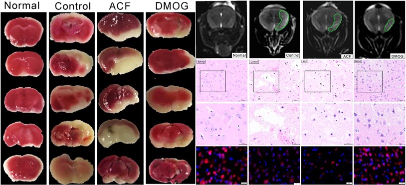 Fig. 2 The results of TTC staining, T2W Magnetic resonance image, H&E staining, and NeuN immunostaining.2
References
- Maida, Carlo Domenico et al. "Molecular Pathogenesis of Ischemic and Hemorrhagic Strokes: Background and Therapeutic Approaches." International Journal of Molecular Sciences vol. 25,12 6297. Distributed under Open Access license CC BY 4.0, without modification. https://doi.org/10.3390/ijms25126297
- Amin, Nashwa et al. "Hypoxia Inducible Factor-1α Attenuates Ischemic Brain Damage by Modulating Inflammatory Response and Glial Activity." Cells vol. 10,6 1359. Distributed under Open Access license CC BY 4.0, with modification. https://doi.org/10.3390/cells10061359
For Research Use Only.