
Calvarial Defect Modeling & Pharmacodynamics Service
Are you currently facing challenges such as slow translational timelines, inconsistent spontaneous healing in preclinical models, or difficulties in quantifying the true osteoinductive potential of your novel biomaterials? Our Calvarial Defect Modeling & Pharmacodynamics Service helps you obtain definitive, high-resolution efficacy data and accelerate your regulatory submission process through our validated critical-size defect (CSD) protocols and multi-dimensional imaging analysis. We utilize specialized surgical techniques and advanced Micro-CT analytics to ensure your therapeutic candidates are evaluated with the highest scientific rigor under 100 words.
Overview of Calvarial Defect Modeling & Pharmacodynamics
The calvaria, or skullcap, serves as a vital physiological system for studying intramembranous ossification, the same process responsible for the development of craniofacial bones in humans. Unlike long bones, which heal via a cartilage intermediate, calvarial healing provides a direct window into osteoblast activity and mineralization. Disruptions in this system due to trauma, congenital deformities (such as craniosynostosis), or oncological resections lead to non-healing bone voids. Our services simulate these pathological states to test the efficacy of scaffolds, growth factors, and cell-based interventions designed to restore structural integrity to the skeletal system. Developing bone substitutes requires models that strictly prevent spontaneous healing to avoid false-positive results. Precise calvarial modeling is essential to provide the mechanical and physiological environment needed to validate xenografts and cell-based therapies.
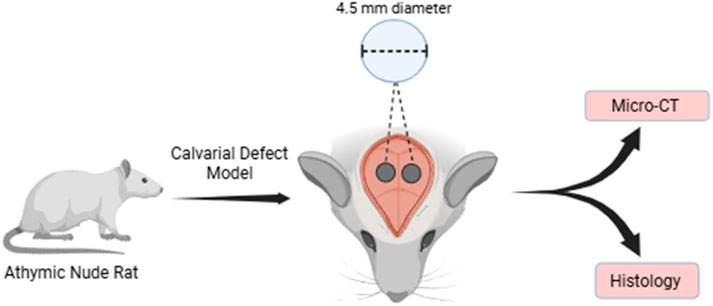 Fig.1 Schematic of calvarial defect model design.1
Calvarial Defect Models
Creative Biolabs provides validated Rat and Rabbit models to support diverse R&D requirements. We specialize in the Critical-Size Defect (CSD), defined as the smallest intraosseous wound that will not heal spontaneously. By precisely controlling the defect diameter (e.g., 4.5 mm to 8 mm depending on species), we ensure that any bone formation observed is strictly a result of the administered treatment. These models are the gold standard for evaluating osteoconduction, osteoinduction, and the degradation kinetics of synthetic biomaterials under standardized conditions.
Evaluation Platform of Our Service
Our platform offers a comprehensive suite of technical parameters to quantify every aspect of the bone healing cascade:
-
Biochemical & Molecular Analysis:
- Cytokine assays (IL-1β, IL-6, TNF-α) to assess local inflammatory response.
- Western Blot for protein expression of bone markers (Runx2, OCN).
- qPCR for gene expression related to osteogenesis and angiogenesis.
-
Histopathological Examination:
- Standardized staining: H&E, Masson's Trichrome, and Goldner's Trichrome.
- Mineralization visualization: Von Kossa and Alizarin Red staining.
- Immunohistochemistry (IHC): BMP-2, VEGF, and Osteocalcin localization.
-
Behavioral & Functional Testing:
- Post-operative pain assessment and activity monitoring.
- Neurological safety screening for craniofacial interventions.
-
Advanced Imaging & Instrumentation:
- High-resolution Micro-CT: BV/TV, Tb.N, Tb.Th, and Tb.Sp measurements.
- Bone Mineral Density (BMD) quantification.
- 3D reconstruction of defect bridging and scaffold resorption.
Key Applications
Our models simulate a wide range of clinical indications, including craniofacial trauma, alveolar bone loss, and post-tumor resection defects. We provide a robust platform to evaluate:
- Small Molecules: Osteogenic stimulants and anti-inflammatory compounds.
- Biologics: Recombinant growth factors and targeted antibodies.
- Gene Therapies: Viral and non-viral vectors promoting localized bone growth.
- Cell-Based Therapies: Seeded scaffolds utilizing Mesenchymal Stem Cells (MSCs).
Why Choose Us?
Wide range of validated Rat and Rabbit strains, including athymic models for xenotransplantation.
Seamless transition from initial in vitro biocompatibility screening to full-scale in vivo PD/PK studies.
PhD-level team with rigorous quality management systems and over 20 years of experience in translational bone research.
Work with Us
- Summarize the project requirements and fill in the information collection form.
- Sign a CDA from both parties to further communicate information, such as targets.
- Select an animal model, discuss experimental design, and determine assay parameters.
- Project costing and project schedule forecasting.
- We provide a detailed project plan, including the required sample quantities, methods, and protocols.
- Both parties confirm the project details and start the project.
- Confirm the timeline of the project.
- We provide periodic results and information on the animal's condition.
- We will work together to make project adjustments as necessary.
- We provide a comprehensive project report promptly.
- We arrange transportation for the produced samples.
- We provide a discussion of the project results and help to arrange the next steps.
- Data storage and archiving.
Frequently Asked Questions
-
Q: How do you ensure the defect in the rat model doesn't heal spontaneously?
A: We utilize a verified 4.5 mm threshold in our rat models. Our longitudinal data shows that spontaneous healing remains below 7-10% over 8 weeks, providing a clear window to observe your test agent's efficacy.
-
Q: What is the typical duration of a calvarial efficacy study?
A: While early signaling can be detected within 4 weeks, we generally recommend an 8-to-12-week study duration to capture definitive mineralization and architectural remodeling.
-
Q: How do you measure the quality of the new bone, not just the volume?
A: Our Micro-CT platform calculates trabecular thickness and connectivity, while histological scoring evaluates the maturity of the lamellar bone compared to woven bone.
-
Q: Is it possible to test liquid or injectable formulations?
A: Yes. Our surgeons are trained in various application techniques, including the use of localized membranes or gels to ensure the test agent remains within the defect site.
Published Data
Objective: To establish a definitive "zero-growth" baseline for critical-size calvarial defects in an immunodeficient model for human-cell therapy validation.
Model Used: Athymic Rat Calvarial Defect Model (4.5 mm diameter).
Results: Micro-CT and histological analysis at 8 weeks post-operation demonstrated a spontaneous bone volume fraction (BV/TV) of less than 7.5%. Histology confirmed the presence of only loose fibrous tissue in the central defect area, with no evidence of bridging or significant mineralization. This study successfully validated the 4.5 mm threshold as a true CSD in athymic rats, providing a reliable platform for evaluating highly potent osteoinductive agents.
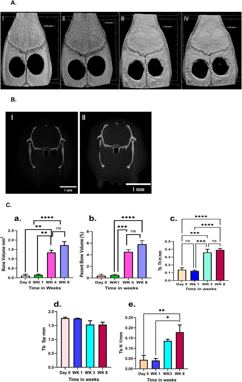 Fig.2 Micro-CT data illustrating the defect group.1
Creative Biolabs is dedicated to providing the most precise and reproducible calvarial defect models in the industry. Whether you are validating a novel 3D-printed scaffold or a complex gene therapy, our team ensures your data is robust, quantitative, and ready for regulatory submission. Contact our specialists, who are standing by to provide technical consultations and tailored study designs.
Reference
- Parco, Todd M., et al. "Comprehensive evaluation of critical-size calvarial defect in athymic rat model." Frontiers in Physiology 16 (2025): 1662424. Distributed under Open Access license CC BY 4.0. The image has been modified; only part of the original image. DOI: https://doi.org/10.3389/fphys.2025.1662424.
For Research Use Only.