
Ovariectomy (OVX) induced Osteoporosis Modeling & Pharmacodynamics Service
Are you currently facing high variability in bone density data, inconsistent surgical outcomes, or challenges in translating preclinical results to human clinical trials? Our Ovariectomy (OVX)-Induced Osteoporosis Modeling & Pharmacodynamics Service helps you obtain high-quality, reproducible efficacy data and accelerate your drug development timeline through our standardized surgical protocols, aged-animal modeling expertise, and multi-dimensional histomorphometric analysis platforms. Under the guidance of our PhD-led team, Creative Biolabs transforms complex biological challenges into actionable scientific insights.
Overview of OVX-Induced Osteoporosis
The skeletal system is a dynamic tissue regulated by the delicate balance between osteoclast-mediated resorption and osteoblast-mediated formation. Postmenopausal osteoporosis is primarily driven by the cessation of ovarian function, leading to a sharp decline in systemic estrogen levels. This hormonal withdrawal triggers a pro-inflammatory cytokine cascade, specifically involving IL-1β, IL-6, and TNF-α, which accelerates osteoclastogenesis and deteriorates the bone microarchitecture. Ovariectomy (OVX) in female animals effectively mimics this physiological shift, providing a robust proxy for human postmenopausal bone loss, characterized by decreased bone mineral density (BMD) and increased fracture risk.
OVX-Induced Osteoporosis Model
Creative Biolabs offers highly standardized OVX models utilizing the Rat and Rabbit species to facilitate diverse research requirements. Our protocols emphasize the use of skeletally mature animals to ensure that bone loss is a result of estrogen deficiency rather than growth-related modeling. We provide both midline and dorsolateral surgical approaches, verified through post-operative uterine atrophy measurements and longitudinal BMD tracking. These models are ideal for evaluating long-term structural integrity and the mechanical performance of new therapeutic agents.
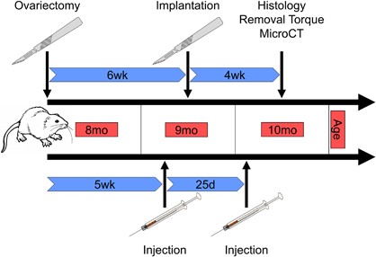 Fig.1 Procedure of drug administration pre- and post-implantation in ovariectomized osteoporotic aged rats.1
Evaluation Platform of Our Service
Our multi-dimensional analysis platform provides a granular view of bone health, ranging from molecular signaling to macro-mechanical strength. We deliver a comprehensive technical parameter list for every study:
-
Biochemical & Molecular Analysis:
- Serum Biomarkers: Estradiol (E2), Osteocalcin (BGP), Alkaline Phosphatase (ALP).
- Bone Turnover Markers: P1NP (Formation), CTX-I (Resorption).
- Cytokine Profiling: IL-1β, IL-6, TNF-α, and Cox-2 mRNA/protein expression via qPCR and Western Blot.
-
Histopathological Examination:
- Undecalcified Bone Sectioning: Goldner's Trichrome and Von Gieson's staining.
- Dynamic Histomorphometry: Mineral Apposition Rate and Bone Formation Rate (BFR).
- Immunohistochemistry (IHC): RANKL/OPG ratio and sclerostin expression.
-
Behavioral & Functional Testing:
- Mechanical Loading Response: Gait analysis and weight-bearing distribution.
- Physical Performance: Grip strength testing for musculoskeletal synergy evaluation.
-
Advanced Imaging & Instrumentation:
- Micro-CT Analysis: 3D quantification of BV/TV, Tb.N, Tb.Th, and Tb.Sp.
- Dual-Energy X-ray Absorptiometry (DEXA): Longitudinal Bone Mineral Density (BMD) measurement.
- Biomechanical Testing: Three-point bending, femoral neck load-to-failure, and removal torque testing for implants.
Key Applications
Our OVX models are optimized for simulating a wide range of clinical indications and therapeutic modalities:
- Indications: Postmenopausal osteoporosis, implant osseointegration in osteoporotic bone, estrogen-deficiency-induced sarcopenia, and fracture healing under metabolic stress.
- Therapeutic Evaluation: Small molecule inhibitors, biologics (monoclonal antibodies), gene therapies targeting bone-anabolic pathways, and advanced cell-based regenerative therapies.
Why Choose Us?
We provide validated, high-health status Rat and Rabbit strains, ensuring the right physiological fit for your specific molecule size and surgical requirements.
Our workflow is seamless, transitioning your project from initial in vitro mechanism-of-action studies to rigorous in vivo PD/PK and safety assessments.
Your project is managed by a PhD-level team with deep expertise in bone histomorphometry, supported by a rigorous quality management system that ensures data integrity for regulatory filings.
Work with Us
- Summarize the project requirements and fill in the information collection form.
- Sign a CDA from both parties to further communicate information, such as targets.
- Select an animal model, discuss experimental design, and determine assay parameters.
- Project costing and project schedule forecasting.
- We provide a detailed project plan, including the required sample quantities, methods, and protocols.
- Both parties confirm the project details and start the project.
- Confirm the timeline of the project.
- We provide periodic results and information on the animal's condition.
- We will work together to make project adjustments as necessary.
- We provide a comprehensive project report promptly.
- We arrange transportation for the produced samples.
- We provide a discussion of the project results and help to arrange the next steps.
- Data storage and archiving.
Frequently Asked Questions
-
Q: Why do you recommend 6-month-old rats instead of younger, more cost-effective options?
A: Skeletal maturity is reached at approximately 6 months. Using younger rats can lead to "false" density data as the animals are still in an active modeling phase. We prioritize 6-month-old models to ensure your results reflect true pathological bone loss.
-
Q: Can we evaluate the effect of our drug on dental or orthopedic implant integration?
A: Absolutely. We offer specialized removal torque testing and BIC (Bone-to-Implant Contact) analysis in our OVX models to evaluate how therapeutics influence osseointegration.
-
Q: How do you verify the success of the ovariectomy?
A: We verify every surgery through a combination of serum estradiol monitoring and post-study uterine weight measurement, which is a definitive biological marker of successful estrogen depletion.
-
Q: What is the typical turnaround time for a standard efficacy study?
A: While bone remodeling takes time, early markers can be detected in as little as 14-30 days. Contact us for a detailed timeline tailored to your specific compound.
Published Data
Objective: To evaluate the effect of systemic bisphosphonate treatment (Ibandronate) on implant osseointegration in a postmenopausal model.
Model Used: 8-month-old rat (Ovariectomized).
Results: The study demonstrated that while Ibandronate successfully increased systemic Bone Volume (BV/TV), it did not significantly improve Bone-to-Implant Contact (BIC). Furthermore, biomechanical testing revealed that the high-turnover osteoporotic environment, even when treated, resulted in lower removal torque compared to healthy controls. This highlights the critical need for therapeutics that support functional remodeling rather than just increasing mineral volume.
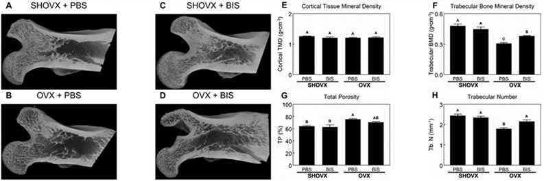 Fig.2 Profile of osteoporotic phenotype induced by ovariectomy.1
Creative Biolabs provides industry-leading Ovariectomy (OVX) modeling and pharmacodynamics services, specializing in Rat and Rabbit research. From high-resolution Micro-CT imaging to complex biomechanical failure testing, we deliver the data necessary to move your candidate from the lab to the clinic with confidence. For detailed project discussions, customized protocols, or pricing information, please reach out to our technical consulting team.
Reference
- Lotz, Ethan M., et al. "Ibandronate treatment before and after implant insertion impairs osseointegration in aged rats with ovariectomy induced osteoporosis." Journal of Bone and Mineral Research Plus 3.7 (2019): e10184. Distributed under Open Access license CC BY 4.0. The image has been modified; only part of the original image. DOI: https://doi.org/10.1002/jbm4.10184.
For Research Use Only.