
Destabilization of the Medial Meniscus (DMM) induced Osteoarthritis Modeling & Pharmacodynamics Service
Are you currently facing challenges in reproducing clinical OA pathology, inconsistent pain behavior data, or high variability in histopathological scoring during preclinical trials? Our DMM-Induced Osteoarthritis Modeling & Pharmacodynamics Service helps you evaluate the efficacy of DMOADs with high sensitivity and reproducibility through our advanced microsurgical platforms, validated OARSI scoring systems, and multi-dimensional behavioral analysis in Rat and Rabbit models.
Overview of DMM-Induced Osteoarthritis
The Medial Meniscal Tear (MMT) model is a premier surgical platform used to simulate post-traumatic osteoarthritis (PTOA). The knee joint is a complex physiological system where the meniscus acts as a vital shock absorber, distributing mechanical loads across the articular cartilage. In the MMT model, a precise transection of the medial meniscus is performed, leading to joint instability and altered biomechanics. This instability triggers a cascade of degenerative events: proteoglycan loss, chondrocyte apoptosis, and eventual full-thickness cartilage erosion. Unlike chemical models that cause immediate metabolic cell death, the MMT model replicates the slow, progressive nature of human OA, making it the "gold standard" for evaluating Disease-Modifying Osteoarthritis Drugs (DMOADs) that aim to protect joint structure or alleviate chronic mechanical pain.
DMM-Induced Disease Models
Osteoarthritis (OA) is a chronic, degenerative joint disease characterized by the progressive breakdown of articular cartilage, subchondral bone remodeling, osteophyte formation, and synovial inflammation. It remains a leading cause of disability worldwide, with a complex etiology involving mechanical stress, metabolic changes, and inflammatory signaling. The Destabilization of the Medial Meniscus (DMM) model has emerged as the premier surgically induced method for simulating human post-traumatic OA. By selectively transecting the medial meniscotibial ligament (MMTL), the meniscus is displaced, leading to an immediate shift in biomechanical loading. This focus on mechanical instability ensures that the resulting cartilage erosion mimics the slow-progression, weight-bearing-dependent pathology observed in human patients, making it superior to intra-articular chemical injections for evaluating long-term disease-modifying agents.
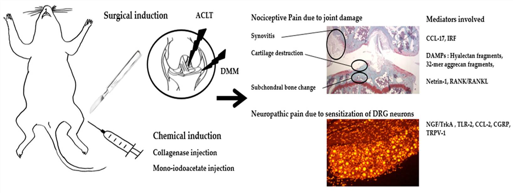 Fig.1 Overview of Molecular Pain Mediators in an Animal Model of Osteoarthritis.1,3
Evaluation Platform of Our Service
Creative Biolabs provides a deep technical parameters list to ensure every facet of joint health and therapeutic response is quantified.
-
Biochemical & Molecular Analysis
- Cytokine Profiles: Quantification via multiplex ELISA.
- Enzymatic Activity: Detect enzyme expression levels.
- Gene Expression: qPCR analysis of inflammatory markers.
- Western Blot: Detection of receptor signaling components.
-
Histopathological Examination
- Standard Staining: Safranin O/Fast Green and H&E.
- Scoring Systems: Validated OARSI (Osteoarthritis Research Society International) histopathology grading.
- Immunohistochemistry (IHC): Spatial mapping of Type II Collagen and MMP-13 degradation fragments.
-
Behavioral & Functional Testing
- Static Weight-Bearing: Analysis of limb-specific incapacitance using advanced pressure sensors.
- Pain Thresholds: von Frey filament testing for mechanical allodynia.
- Gait Analysis: Automated quantification of stride length, swing time, and stance patterns.
-
Advanced Imaging & Instrumentation
- Micro-CT: High-resolution 3D reconstruction of the subchondral bone.
- Bone Mineral Density (BMD): Quantification of subchondral bone sclerosis.
- Morphometric Analysis: Measurement of trabecular thickness (Tb.Th) and bone volume fraction (BV/TV).
Key Applications
Our DMM models are designed to simulate clinical indications, including:
- Post-Traumatic Osteoarthritis (PTOA)
- Age-Related Joint Degeneration
- Subchondral Bone Sclerosis & Osteophyte Pathologies
These platforms are suitable for the evaluation of diverse therapeutic modalities, including small molecule inhibitors, biologics (mAbs, growth factors), gene therapies, and cell-based regenerative medicines.
Why Choose Us?
Our PhD-level surgical team utilizes stereomicroscope-aided DMM induction, significantly reducing the Coefficient of Variation (CV) in pain behavior and OARSI scores compared to traditional "naked-eye" surgeries.
We provide highly validated strains for both Rat and Rabbit models, allowing for scaling from pilot studies to complex pharmacological investigations.
Creative Biolabs offers a seamless transition from initial in vitro chondrocyte screening to comprehensive in vivo PD/PK studies, all under a rigorous quality management system.
Our studies are guided by senior scientists with expertise in the TGF-β and BMP signaling pathways, ensuring your data is interpreted within the context of the latest orthopedic research.
Work with Us
- Summarize the project requirements and fill in the information collection form.
- Sign a CDA from both parties to further communicate information, such as targets.
- Select an animal model, discuss experimental design, and determine assay parameters.
- Project costing and project schedule forecasting.
- We provide a detailed project plan, including the required sample quantities, methods, and protocols.
- Both parties confirm the project details and start the project.
- Confirm the timeline of the project.
- We provide periodic results and information on the animal's condition.
- We will work together to make project adjustments as necessary.
- We provide a comprehensive project report promptly.
- We arrange transportation for the produced samples.
- We provide a discussion of the project results and help to arrange the next steps.
- Data storage and archiving.
Frequently Asked Questions
-
Q: How does the DMM model compare to the ACLT (Anterior Cruciate Ligament Transection) model?
A: While ACLT induces rapid, severe OA, the DMM model is preferred for its slower, more controlled progression. This provides a wider therapeutic window to observe the subtle efficacy of disease-modifying drugs, particularly those targeting early-to-mid stage cartilage preservation.
-
Q: Can you perform DMM in rabbits as well as rats?
A: Yes, Creative Biolabs provides validated DMM protocols for both rats and rabbits. The Rabbit model is particularly useful for studies requiring larger synovial fluid volumes or more extensive imaging of osteophyte formation.
-
Q: What is the typical duration of a DMM pharmacodynamics study?
A: Most studies run for 8 to 12 weeks post-surgery to allow for significant histological changes and behavioral deficits to manifest. However, we can customize time points based on your specific therapeutic mechanism.
-
Q: How do you ensure the reproducibility of pain behavior data?
A: We use stereomicroscope-aided surgery to minimize non-specific inflammation and ensure consistent ligament transection. Combined with standardized environmental acclimation for the animals, this reduces variability in weight-bearing and gait analysis.
Published Data
Objective: To investigate the chondroprotective effects of a Traditional Chinese Medicine (TCM) extract via the TGF-β/Smad signaling pathway in a mouse OA model.
Model Used: DMM-induced OA in C57BL/6 mice (Validation relevant to our Rat/Rabbit platform capabilities).
Results: The study demonstrated that the therapeutic agent successfully ameliorated OA phenotypes, including cartilage degradation and subchondral bone sclerosis. Molecular analysis revealed up-regulation of Col2, TGFβRII, and pSmad-2, while catabolic markers like MMP-13 were significantly down-regulated. The study concluded that the agent decelerated cartilage degradation partly via the TGF-β signaling pathway, highlighting the sensitivity of the DMM model in detecting molecular-level therapeutic shifts.
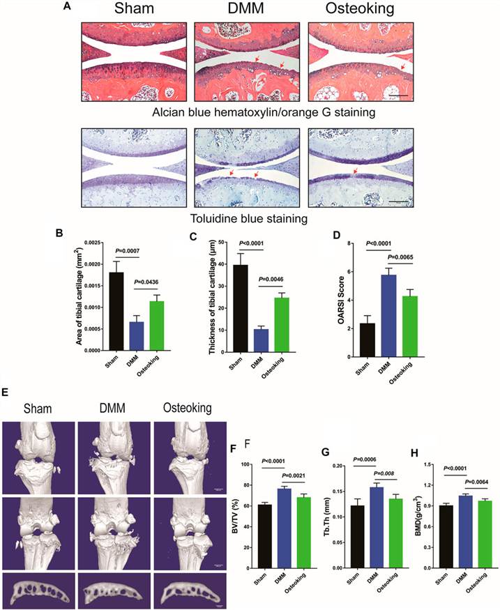 Fig.2 Osteoking slows osteoarthritis progression in a DMM-induced model.2,3
Creative Biolabs is committed to providing the most reliable and clinically relevant DMM-induced OA models to the global biopharmaceutical community. Our integration of microsurgical precision with advanced molecular and behavioral analysis ensures that your drug discovery pipeline is built on a foundation of scientific excellence. Contact our expert team, which is available to provide detailed technical consultations and customized study designs.
References
- Hong, Jeong-Im, In Young Park, and Hyun Ah Kim. "Understanding the molecular mechanisms underlying the pathogenesis of arthritis pain using animal models." International Journal of Molecular Sciences 21.2 (2020): 533. DOI: https://doi.org/10.3390/ijms21020533.
- Ling, Houfu, et al. "Osteoking decelerates cartilage degeneration in DMM-induced osteoarthritic mice model through TGF-β/smad-dependent manner." Frontiers in Pharmacology 12 (2021): 678810. DOI: https://doi.org/10.3389/fphar.2021.678810.
- Distributed under Open Access license CC BY 4.0, without modification.
For Research Use Only.