
Traumatic Brain Injury Modeling & Pharmacodynamics Services
Introduction
Traumatic Brain Injury (TBI) is a leading cause of global mortality and long-term disability, particularly within industrialized nations. According to recent epidemiological data, over 50 million people worldwide suffer a TBI each year, with many survivors facing permanent impairments in cognitive, learning, and motor functions. The complexity of TBI stems from its dual-phase pathology. It is characterized by an initial primary injury, the immediate mechanical disruption of neural tissue, followed by a prolonged secondary injury cascade. This secondary phase involves a devastating cycle of oxidative stress, profound neuroinflammation, and blood-brain barrier (BBB) breakdown. If left unchecked, these processes lead to chronic cognitive deficits and significantly increase the risk of developing Chronic Traumatic Encephalopathy (CTE). Establishing effective therapeutic strategies remains a significant challenge due to the immense variety of clinical presentations. To bridge this gap, Creative Biolabs' platform provides clinically relevant TBI models, ranging from focal contusions to diffuse axonal injuries. By combining these high-fidelity models with multi-modal readouts, we offer a specialized environment to evaluate the efficacy of next-generation neuroprotective agents, anti-inflammatory therapies, and regenerative medicines.
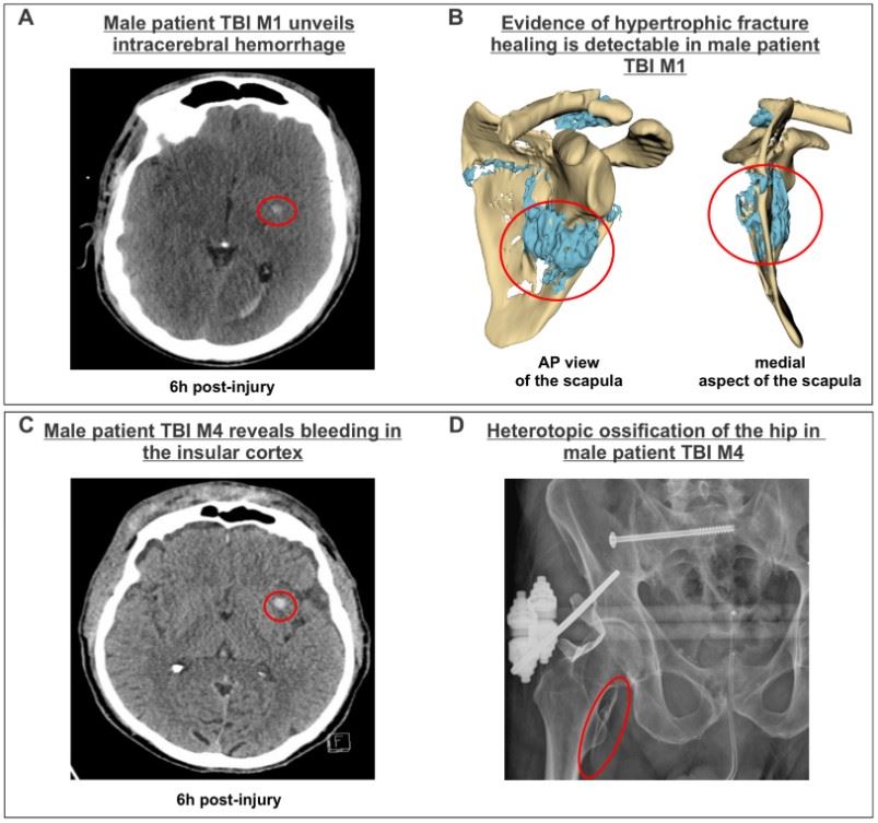 Fig.1 Radiological analysis of male TBI patients demonstrates that acute intracranial hemorrhages in regions such as the centrum semiovale and insular cortex are associated with subsequent musculoskeletal complications, including hypertrophic fracture healing and the development of heterotopic ossification.1
Available Traumatic Brain Injury Models
We provide a high-precision Traumatic Brain Injury (TBI) research platform, including Rodent TBI models and Primate Peripheral Nerve Injury (PNI) Model, to simulate the full clinical spectrum of focal contusions and diffuse axonal injuries. By bridging specialized modeling with translational pharmacodynamics, we empower partners to accelerate the development of neuroprotectants, anti-epileptogenic agents, and neuroinflammatory modulators, to drive lead optimization and preclinical research for both acute trauma and chronic neurodegenerative sequelae.
| TBI Models | Modeling Methods | Application Values | Animal Species |
| Fluid Percussion Injury (FPI) Model | An electromagnetic or pneumatic piston delivers a precise, high-velocity impact to the exposed dura. | Focal Injury & Contusion: The gold standard for replicating focal contusion, intraparenchymal hemorrhage, and cortical loss. Ideal for evaluating Neuroprotective Agents (e.g., Progesterone, Cyclosporine A) and BBB Stabilizers aimed at reducing lesion volume and tissue sparing. | Mouse, Rat |
| Controlled Cortical Impact Injury (CCI) Model | A rapid fluid-pressure pulse is delivered to the intact dura through a fluid-filled reservoir. | Mixed Focal/Diffuse Injury: Replicates subcortical damage and brainstem strain. A premier platform for testing Anti-Epileptic Drugs (AEDs) (e.g., Levetiracetam) and Anti-Inflammatory Modulators (e.g., Minocycline) to prevent post-traumatic epilepsy and chronic gliosis. | Mouse, Rat |
| Weight Drop Model | A calibrated weight is dropped from a specific height onto a helmeted or unhelmeted skull. | Diffuse Axonal Injury (DAI): Simulates blunt force trauma or falls. Ideal for screening Axonal Repair Therapies (e.g., Taxol derivatives), metabolic enhancers, and cognitive boosters (e.g., Donepezil) targeting widespread white matter disruption. | Mouse, Rat |
Evaluation Platform
Our platform integrates structural, functional, and molecular endpoints to provide a 360-degree view of TBI progression.
-
Neuroimaging & Physiological Monitoring:
- 7T Small Animal MRI (T2/DTI): Utilizing Diffusion Tensor Imaging (DTI) to quantify white matter integrity and fractional anisotropy (FA).
- Intracranial Pressure (ICP) Monitoring: Real-time tracking of post-traumatic edema and pressure spikes.
-
Behavioral & Cognitive Battery:
- Morris Water Maze (MWM) / Barnes Maze: Standardized tests for spatial learning and memory deficits associated with hippocampal damage.
- Rotarod & Beam Balance: Quantifying motor coordination and vestibular dysfunction.
- Novel Object Recognition (NOR): Assessing recognition memory and cortical integrity.
-
Molecular & Histopathology:
- BBB Permeability: Quantifying Evans Blue or Fluorescein extravasation.
- Neuroinflammation: IHC for Iba1 (microglia) and GFAP (astrocytes) to map the inflammatory response and glial scar formation.
- Axonal Injury Markers: Quantification of Silver Staining or Amyloid Precursor Protein (APP) accumulation to identify axonal transport disruption.
Applications
- Neuroprotection & Anti-Apoptosis: Evaluating pharmacological agents that mitigate primary mechanical insults and inhibit secondary apoptotic cascades, including excitotoxicity and mitochondrial dysfunction, to preserve the perilesional cortex.
- Anti-Epileptogenesis Screening: Utilizing longitudinal EEG and behavioral monitoring to validate therapies aimed at preventing Post-Traumatic Epilepsy (PTE) and mitigating chronic seizure development following FPI or CCI injury.
- DAI & White Matter Repair: Assessing the efficacy of growth factors and microtubule stabilizers in restoring axonal transport and structural integrity across white matter tracts to reverse widespread connectivity deficits.
- Neuroinflammation & Edema Management: Quantifying the impact of biologics or small molecules on M1/M2 microglial polarization, glial scarring, and tight junction stability to preserve BBB integrity and reduce cerebral edema.
- Chronic Sequelae & Neurodegeneration: Studying the long-term pathological link between TBI and tauopathy, investigating mechanisms that drive Alzheimer's-like neurodegeneration and CTE.
- Translational Functional Recovery: Correlating histological tissue sparing with multi-dimensional outcomes, via MWM and Beam Walking, to provide robust data for IND-enabling drug development.
Our Advantages
- Precision Engineering & Model Fidelity: Using advanced electromagnetic impactors (e.g., Leica Impact One), we achieve sub-millimeter control over depth and velocity to eliminate intra-group variability. Every model is validated by acute clinical markers to ensure injury severity aligns perfectly with your study goals.
- Advanced White Matter & Axonal Assessment: We utilize 7T Small Animal MRI (DTI sequences) to non-invasively track white matter integrity via Fractional Anisotropy. This is complemented by specialized silver staining and APP mapping to provide definitive histological proof of axonal preservation and neural circuit repair.
- Multi-Phase Pharmacodynamic Readouts: Our platform monitors the full pathological spectrum: from acute ICP and BBB permeability to secondary neuroinflammatory cascades (M1/M2 polarization) and chronic outcomes like hippocampal-dependent memory loss and Tau pathology.
- Ultra-Sensitive Biomarker Integration: Using SIMOA/MSD platforms, we quantify gold-standard biomarkers (e.g., Neurofilament Light (NfL), GFAP, UCH-L1) in plasma and CSF. This allows you to correlate preclinical drug efficacy with the exact same endpoints used in human clinical trials.
- Specialized Delivery & Surgical Expertise: We offer expert intraparenchymal, Intracerebroventricular (ICV), and osmotic pump delivery to ensure your candidate bypasses the BBB effectively. Our team can pivot between focal (CCI) and diffuse (Impact Acceleration) models to match your drug's specific mechanism of action.
Work with Us
- Summarize the project requirements and fill in the information collection form.
- Sign a CDA from both parties to further communicate information, such as targets.
- Select an animal model, discuss experimental design, and determine assay parameters.
- Project costing and project schedule forecasting.
- We provide a detailed project plan, including the required sample quantities, methods, and protocols.
- Both parties confirm the project details and start the project.
- Confirm the timeline of the project.
- We provide periodic results and information on the animal's condition.
- We will work together to make project adjustments as necessary.
- We provide a comprehensive project report promptly.
- We arrange transportation for the produced samples.
- We provide a discussion of the project results and help to arrange the next steps.
- Data storage and archiving.
FAQs
-
Q: How do you choose between the CCI and FPI models?
A: The choice depends on your therapeutic target. CCI is best for focal injury research. It produces a highly reproducible cortical contusion and is ideal for studying cell death, focal neuroinflammation, and cortical tissue sparing. FPI is better for mixed focal/diffuse injury. It creates a pressure wave that affects subcortical structures (like the hippocampus and brainstem). It is the preferred model for studying post-traumatic epilepsy and concussive-like symptoms.
-
Q: How do you ensure the mechanical impact is consistent across a study cohort?
A: We use computerized electromagnetic impactors (e.g., Leica Impact One) that allow for sub-millimeter precision. For every animal, we record and report the exact impact velocity, dwell time, and depth. Any animal where the impact parameters deviate by more than 5% is excluded to ensure the resulting pharmacodynamic data is robust and statistically sound.
-
Q: Can your platform model "Mild TBI" or Concussion without a craniotomy?
A: Yes. We offer Closed Head Injury (CHI) and Impact Acceleration models. These involve an impact to the intact skull (often protected by a helmet/disk), which replicates the DAI and metabolic crisis seen in human concussions without the focal cavitation caused by direct cortical impact.
-
Q: What is the "Righting Reflex" and why is it measured?
A: The Righting Reflex Time (RRT) is the time it takes for an animal to flip from its back to its feet following injury. It serves as an immediate surrogate for the "duration of loss of consciousness" (LOC). It is a vital acute endpoint to confirm injury severity and can be used to screen neuroprotective agents that aim to reduce acute neurological suppression.
-
Q: How do you quantify BBB disruption?
A: We primarily use Evans Blue or Sodium Fluorescein extravasation assays. The dye is injected systemically and, in the event of BBB breakdown, leaks into the brain parenchyma. We then perform spectrophotometric quantification or fluorescence imaging of brain sections to determine the degree of vascular permeability.
-
Q: Which biomarkers are most reliable for tracking TBI progression in rodents?
A: We utilize ultra-sensitive SIMOA or MSD platforms to track NfL (A gold-standard marker for axonal injury), GFAP (An indicator of astrogliosis and acute brain damage), and UCH-L1 (A marker of neuronal cell body injury). These can be measured longitudinally in plasma to correlate with behavioral recovery.
-
Q: Can you perform cognitive testing in TBI models?
A: Absolutely. TBI often affects the hippocampus and prefrontal cortex. We use the MWM or Barnes Maze to evaluate spatial learning and memory deficits. We also offer the NOR test to assess higher-order cognitive processing and recognition memory.
Published Data
Progesterone significantly attenuates brain damage in rats subjected to repetitive mild Fluid Percussion Injury (rFPI). This neuroprotective effect is achieved by suppressing neuroinflammation (evidenced by reduced GFAP immunoreactive areas and CD68-positive cells) and mitigating oxidative stress (evidenced by decreased MDA levels). Consequently, progesterone treatment preserves gray and white matter volumes and improves the structural integrity of the corpus callosum, facilitating functional recovery.
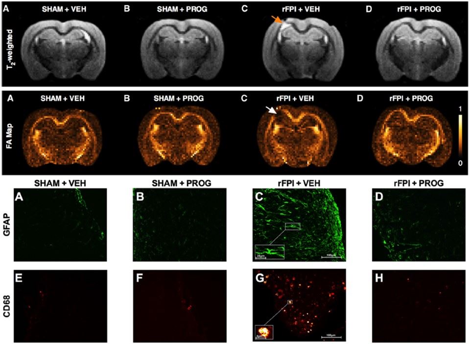 Fig. 2 PROG treatment attenuates brain atrophy, mitigates corpus callosum injury, and reduces neuroinflammation and oxidative stress after rFPI.2
References
- Joneleit, Jonas et al. "A Novel Human Stem Cell Culture Model for Severe Traumatic Brain Injury Reflecting Sexual Dimorphism in Heterotopic Ossification." Cells vol. 14,19 1491. Distributed under Open Access license CC BY 4.0, with modification. https://doi.org/10.3390/cells14191491
- Webster, Kyria M et al. "Progesterone treatment reduces neuroinflammation, oxidative stress and brain damage and improves long-term outcomes in a rat model of repeated mild traumatic brain injury." Journal of neuroinflammation vol. 12 238. Distributed under Open Access license CC BY 4.0, with modification. https://doi.org/10.1186/s12974-015-0457-7
For Research Use Only.