
Destabilization of the Medial Meniscus (DMM) & Anterior Cruciate Ligament Transection (ACLT) induced Osteoarthritis Modeling & Pharmacodynamics Service
Are you currently facing long drug development cycles or difficulty in accurately recapitulating human joint degradation in preclinical stages? Our DMM & ACLT-Induced Osteoarthritis Modeling & Pharmacodynamics Service helps you obtain high-quality, reproducible efficacy data through precision surgical induction and multi-omic validation platforms. We streamline the path from bench to clinic by utilizing advanced histomorphometry and transcriptomic analysis to verify your therapeutic's impact on cartilage and bone health.
Overview of Osteoarthritis Modeling
Osteoarthritis (OA) is a chronic, degenerative disorder of the synovial joints, characterized by the progressive loss of articular cartilage, subchondral bone remodeling, and chronic inflammation. It is a leading cause of disability worldwide, yet current treatments are largely limited to pain management rather than structural repair. Animal models that induce joint instability, specifically via Destabilization of the Medial Meniscus (DMM) or Anterior Cruciate Ligament Transection (ACLT), are critical for simulating the mechanical triggers of human post-traumatic osteoarthritis (PTOA). These models allow researchers to observe the transition from initial joint insult to irreversible structural failure, providing a window for testing Disease-Modifying Osteoarthritis Drugs (DMOADs).
DMM & ACLT-Induced Disease Models
Creative Biolabs provides validated OA models across multiple species, including Rat, Rabbit, Cat, and Dog. Our DMM model involves the precise transection of the medial meniscotibial ligament, offering a slowly progressing, highly reproducible pathology ideal for long-term efficacy studies. For more aggressive degradation, our ACLT model replicates acute traumatic injury, leading to rapid cartilage erosion and subchondral bone changes. Each model is executed by specialist surgeons to minimize iatrogenic damage and maximize the reliability of your pharmacodynamic data.
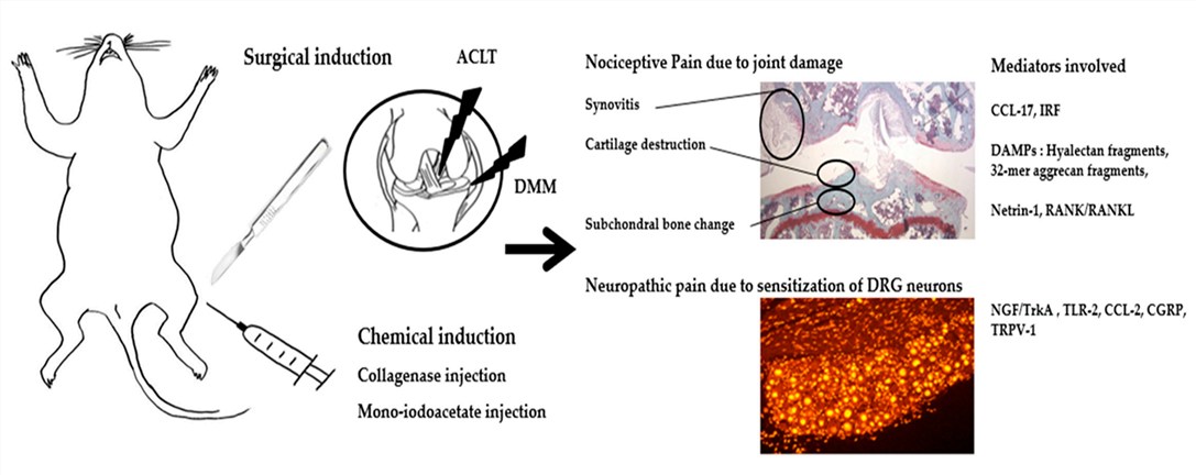 Fig.1 Compilation of molecular pain mediators in osteoarthritis animal model.1,3
Evaluation Platform of Our Service
Creative Biolabs offers a multi-dimensional analysis platform to quantify joint health with surgical precision. Our technical capabilities include:
-
Biochemical & Molecular Analysis:
- Cytokine Profiling: Multiplex assays in synovial fluid and serum.
- Gene Expression: qPCR and transcriptomic profiling (RNA-Seq) for markers.
- Protein Quantification: Western Blot and ELISA for matrix degradation fragments.
-
Histopathological Examination:
- Advanced Staining: Safranin-O/Fast Green and Toluidine Blue for proteoglycan assessment.
- Scoring Systems: OARSI, Glasson, and Mankin grading by board-certified pathologists.
- IHC/IF: Localization of inflammatory markers and chondrocyte viability indicators.
-
Behavioral & Functional Testing:
- Gait Analysis: Automated tracking of limb weight-bearing and stride length.
- Pain Assessment: Von Frey filaments for mechanical allodynia and pressure pain threshold.
- Motor Coordination: Rotarod and grip strength testing for overall joint function.
-
Advanced Imaging & Instrumentation:
- Micro-CT: High-resolution 3D analysis of subchondral bone plate (SBP) thickness and osteophyte volume.
- MRI: Non-invasive longitudinal monitoring of soft tissue and cartilage volume.
- BMD Measurement: Dual-energy X-ray absorptiometry (DEXA) for bone mineral density changes.
Key Applications
Our OA modeling platform is designed to simulate a variety of clinical indications and evaluate diverse therapeutic modalities:
- Simulated Indications: Post-traumatic Osteoarthritis (PTOA), age-related degenerative joint disease, and secondary OA following meniscal injury.
- Small Molecules: Selective kinase inhibitors and anti-inflammatory compounds.
- Biologics: Monoclonal antibodies and growth factors.
- Gene Therapies: Viral vector-mediated delivery of chondroprotective genes.
- Cell-Based Therapies: Mesenchymal stem cell (MSC) injections and tissue-engineered constructs.
Why Choose Us?
Creative Biolabs stands as a leader in musculoskeletal research by combining technical mastery with strategic scientific support.
We offer fully validated models in Rat, Rabbit, Cat, and Dog, allowing you to choose the physiological scale that best fits your therapeutic mechanism of action.
Our platform supports your project from initial in vitro chondrocyte screening through to comprehensive in vivo PD/PK studies, ensuring data consistency across the development cycle.
Projects are managed by a PhD-level team with decades of experience in orthopedic surgery and histopathology. Our rigorous quality management system ensures that every slide and data point is audit-ready.
Work with Us
- Summarize the project requirements and fill in the information collection form.
- Sign a CDA from both parties to further communicate information, such as targets.
- Select an animal model, discuss experimental design, and determine assay parameters.
- Project costing and project schedule forecasting.
- We provide a detailed project plan, including the required sample quantities, methods, and protocols.
- Both parties confirm the project details and start the project.
- Confirm the timeline of the project.
- We provide periodic results and information on the animal's condition.
- We will work together to make project adjustments as necessary.
- We provide a comprehensive project report promptly.
- We arrange transportation for the produced samples.
- We provide a discussion of the project results and help to arrange the next steps.
- Data storage and archiving.
Frequently Asked Questions
-
Q: Can Creative Biolabs perform longitudinal imaging in Rabbit or Dog models?
A: Yes, we utilize high-resolution MRI and Micro-CT for longitudinal monitoring in larger species like rabbits and dogs, allowing you to track structural changes in the same animal over time.
-
Q: How do you ensure surgical reproducibility in the DMM model?
A: Our surgeons utilize specialized micro-instruments and intra-operative magnification. We also include "Sham" groups to ensure that any observed pathology is due to ligament transection rather than the surgical procedure itself.
-
Q: What is the typical lead time for a Rat DMM study?
A: A standard study typically runs for 8 to 12 weeks post-surgery to allow for significant OA development. Contact us for a custom timeline based on your specific requirements.
Published Data
Objective: To compare the transcriptomic response and histological progression between non-surgical injury and surgical DMM models.
Model Used: Surgical DMM and ACL Rupture in comparable cohorts.
Results: Comparative transcriptomic analysis revealed a high correlation (Cosine similarity) between DMM and ACL injury signatures. Key findings included the significant upregulation of miR-199-5p and its involvement in regulating chondrocyte homeostasis. Histological scoring confirmed that DMM provided a reliable, slowly progressing degradation profile, while ACL-induced changes were more rapid, validating both as complementary tools for drug assessment.
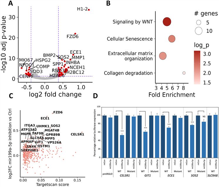 Fig.2 Suppression of miR-199a-5p triggers a catabolic state.2,3
Creative Biolabs provides a premier destination for DMM & ACLT-Induced Osteoarthritis Modeling & Pharmacodynamics. With our expertise in multiple species—Rat, Rabbit, Cat, and Dog—and our advanced multi-omic evaluation platform, we provide the evidence you need to de-risk your clinical pipeline. Contact our team of specialists to discuss your study design, species requirements, and timeline.
References
- Hong, Jeong-Im, In Young Park, and Hyun Ah Kim. "Understanding the molecular mechanisms underlying the pathogenesis of arthritis pain using animal models." International Journal of Molecular Sciences 21.2 (2020): 533. DOI: https://doi.org/10.3390/ijms21020533.
- Gilbert, Sophie J., et al. "Comparative transcriptomic analysis of articular cartilage of post-traumatic osteoarthritis models." Disease Models & Mechanisms 17.10 (2024): dmm050583. DOI: https://doi.org/10.1242/dmm.050583.
- Distributed under Open Access license CC BY 4.0, without modification.
For Research Use Only.