
Monosodium Iodoacetate (MIA) induced Osteoarthritis Modeling & Pharmacodynamics Service
Are you currently facing long drug development cycles, high variability in spontaneous OA models, or challenges in quantifying pain-related behaviors? Our Monosodium Iodoacetate-Induced Osteoarthritis Modeling & Pharmacodynamics Service helps you obtain highly reproducible pathological data and evaluate therapeutic efficacy through precisely controlled chemical induction and multi-dimensional functional analysis. Under our expert guidance, your DMOAD candidates undergo rigorous validation using industry-standard protocols, helping you obtain high-quality data and streamline your path to clinical trials through advanced imaging and innovative histopathological techniques.
Overview of Monosodium Iodoacetate-Induced Osteoarthritis Modeling
Osteoarthritis (OA) is a chronic, degenerative joint disease characterized by the progressive breakdown of articular cartilage, subchondral bone remodeling, and synovial inflammation. It is a leading cause of global disability, primarily affecting weight-bearing joints such as the knee. The physiological system of the joint relies on a delicate balance between chondrocyte metabolism and extracellular matrix (ECM) integrity. In OA, this balance is disrupted, leading to the "undruggable" cycle of inflammation and mechanical failure. Monosodium Iodoacetate (MIA) acts as a metabolic inhibitor that targets glyceraldehyde-3-phosphate dehydrogenase (GAPDH). This action halts glycolysis, inducing chondrocyte apoptosis and rapid cartilage matrix collapse, effectively simulating the metabolic and degenerative hallmarks of human OA in a controlled preclinical setting.
Monosodium Iodoacetate-Induced Osteoarthritis Models
Creative Biolabs provides highly standardized MIA-induced models across multiple species to suit different research objectives. By utilizing intra-articular injections, we bypass the variability of surgical trauma, creating a biphasic disease profile that includes an initial acute inflammatory stage followed by a chronic degenerative phase. Our models are validated in Mouse, Rat, Rabbit, Cat, and Dog, ensuring that whether you are studying early-stage synovial cytokines or late-stage subchondral bone sclerosis, we provide the most relevant physiological environment for your therapeutic candidate.
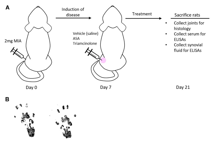 Fig.1 Experimental model of osteoarthritis using rat monosodium iodoacetate (MIA).1,3
Evaluation Platform of Our Service
Creative Biolabs utilizes a multi-dimensional analysis suite to quantify the impact of your therapeutic agents. Our technical parameters include:
-
Biochemical & Molecular Analysis:
- Cytokine Profiling: Multiplex assays.
- Biomarker Quantitation: ELISA.
- Gene Expression: qPCR analysis of cartilage matrix genes.
-
Histopathological Examination:
- Staining Protocols: Safranin-O/Fast Green, H&E, and Toluidine Blue.
- Scoring Systems: Standardized OARSI and Mankin grading.
- Immunohistochemistry (IHC): Detection of Caspase-3, Type II Collagen, and Macrophage markers.
-
Behavioral & Functional Testing:
- Weight-Bearing Analysis: Static incapacitance testing for hindlimb asymmetry.
- Mechanical Allodynia: Von Frey filament testing for pain thresholds.
- Gait Analysis: Automated tracking of stride length and swing time.
-
Advanced Imaging & Instrumentation:
- Micro-CT: Subchondral bone volume/tissue volume (BV/TV) and BMD measurement.
- High-Res MRI: Non-invasive longitudinal monitoring of joint space narrowing.
Key Applications
Our MIA models simulate various clinical indications, including post-traumatic OA, inflammatory joint flares, and chronic neuropathic joint pain. These models are ideal for evaluating a wide spectrum of therapeutic agents:
- Small Molecules: NSAIDs, kinase inhibitors, and metabolic modulators.
- Biologics: Monoclonal antibodies targeting NGF or pro-inflammatory cytokines.
- Gene Therapies: AAV-mediated delivery of chondroprotective factors.
- Cell-Based Therapies: Evaluation of MSC-derived secretomes and regenerative treatments.
Why Choose Us?
We offer validated MIA protocols for Mouse, Rat, Rabbit, Cat, and Dog, ensuring the model matches your specific translational requirements.
Creative Biolabs provides a seamless workflow from initial in vitro screening to comprehensive in vivo PD/PK studies and regulatory-ready data packages.
Our PhD-level team leverages over 20 years of experience to design optimized study protocols, backed by a rigorous quality management system that ensures data integrity and reproducibility.
Workf with Us
- Summarize the project requirements and fill in the information collection form.
- Sign a CDA from both parties to further communicate information, such as targets.
- Select an animal model, discuss experimental design, and determine assay parameters.
- Project costing and project schedule forecasting.
- We provide a detailed project plan, including the required sample quantities, methods, and protocols.
- Both parties confirm the project details and start the project.
- Confirm the timeline of the project.
- We provide periodic results and information on the animal's condition.
- We will work together to make project adjustments as necessary.
- We provide a comprehensive project report promptly.
- We arrange transportation for the produced samples.
- We provide a discussion of the project results and help to arrange the next steps.
- Data storage and archiving.
Frequently Asked Questions
-
Q: How quickly does the MIA model progress compared to surgical models?
A: The MIA model is significantly faster, with structural degradation and pain markers appearing as early as Day 3-7, whereas surgical models may take months to develop similar pathology.
-
Q: Can we test both analgesic and disease-modifying effects in the same study?
A: Yes! The MIA model features a biphasic profile, an early inflammatory phase, and a late degenerative phase, allowing for the assessment of both symptomatic relief and structural preservation.
-
Q: Are the rabbit or dog models suitable for intra-articular injections of viscous gels?
A: Absolutely. The larger joint volumes in rabbits and dogs make them ideal for testing injectable hydrogels, scaffolds, or long-acting release formulations.
-
Q: How does Creative Biolabs ensure the reproducibility of behavioral pain testing?
A: We use standardized, environment-controlled testing rooms and blinded operators to minimize variability and ensure statistically significant behavioral data.
Published Data
Objective: To evaluate the effect of macrophage-derived factors on cartilage preservation and pain reduction in an OA environment.
Model Used: Sprague-Dawley Rat MIA-induced OA model (2 mg intra-articular injection).
Results: This model successfully recapitulated rapid cartilage degradation and neuropathic pain. Micro-CT and OARSI-based histopathology demonstrated that macrophage modulation significantly mitigated cartilage fibrillation and subchondral bone loss. Crucially, incapacitance testing showed a definitive reversal of weight-bearing asymmetry. These results highlight the MIA model's superior sensitivity in capturing the metabolic collapse of the joint and its high fidelity in evaluating drugs targeting the synovial microenvironment and pain-related signaling axes.
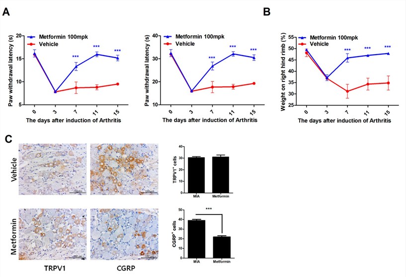 Fig.2 Metformin alleviates pain in rats with MIA-induced osteoarthritis.2,3
Creative Biolabs offers a premier Monosodium Iodoacetate-Induced Osteoarthritis Modeling & Pharmacodynamics Service, providing the technical depth and species versatility (Mouse, Rat, Rabbit, Cat, and Dog) required to move your OA program forward. From precision imaging to complex behavioral metrics, we provide the evidence you need for clinical success. Our expert team is standing by to provide a detailed consultation and customized study design.
References
- Kimmerling, Kelly A., et al. "Amniotic suspension allograft modulates inflammation in a rat pain model of osteoarthritis." Journal of Orthopaedic Research® 38.5 (2020): 1141-1149. DOI: https://doi.org/10.1002%2Fjor.24559.
- Na, Hyun Sik, et al. "Metformin attenuates monosodium-iodoacetate-induced osteoarthritis via regulation of pain mediators and the autophagy–lysosomal pathway." Cells 10.3 (2021): 681. DOI: https://doi.org/10.3390/cells10030681.
- Distributed under Open Access license CC BY 4.0, without modification.
For Research Use Only.